. 2015 Nov;36(11):1070-9.
doi: 10.1002/humu.22833. Epub 2015 Aug 6.

POLD1 Germline Mutations in Patients Initially Diagnosed with Werner Syndrome

Affiliations

POLD1 Germline Mutations in Patients Initially Diagnosed with Werner Syndrome

Davor Lessel et al. Hum Mutat. 2015 Nov.

Abstract

Segmental progeroid syndromes are rare, heterogeneous disorders characterized by signs of premature aging affecting more than one tissue or organ. A prototypic example is the Werner syndrome (WS), caused by biallelic germline mutations in the Werner helicase gene (WRN). While heterozygous lamin A/C (LMNA) mutations are found in a few nonclassical cases of WS, another 10%-15% of patients initially diagnosed with WS do not have mutations in WRN or LMNA. Germline POLD1 mutations were recently reported in five patients with another segmental progeroid disorder: mandibular hypoplasia, deafness, progeroid features syndrome. Here, we describe eight additional patients with heterozygous POLD1 mutations, thereby substantially expanding the characterization of this new example of segmental progeroid disorders. First, we identified POLD1 mutations in patients initially diagnosed with WS. Second, we describe POLD1 mutation carriers without clinically relevant hearing impairment or mandibular underdevelopment, both previously thought to represent obligate diagnostic features. These patients also exhibit a lower incidence of metabolic abnormalities and joint contractures. Third, we document postnatal short stature and premature greying/loss of hair in POLD1 mutation carriers. We conclude that POLD1 germline mutations can result in a variably expressed and probably underdiagnosed segmental progeroid syndrome.

Keywords: POLD1; Werner syndrome; deafness; mandibular hypoplasia; progeroid features (MDP) syndrome.


Figures

Figure 1
Figure 1
Identification of causative POLD1 mutations. A: Protein structure of POLD1. The position of the mutations identified in progeroid patients are marked with vertical arrows and shown in black; cancer-associated mutations are shown in red. (NN terminus; C-C terminus). B: Sanger sequencing chromatograms of parts of POLD1 exon 13 after PCR amplification of genomic DNA. Sequencing results for MINN1010 bearing a heterozygous c.1519C>T (p.R507C) and an individual homozygous for the reference sequence (WT/WT). The amino acid translation is shown in the three-letter code above the chromatograms. C: Sanger sequencing chromatograms of parts of POLD1 exon 15 after PCR amplification of genomic DNA. Sequencing results for HD1010 bearing a heterozygous c.1812_1814delCTC (p.Ser605del) and an individual homozygous for the reference sequence (WT/WT). The amino acid translation is shown in the three letter code above the chromatograms. Nucleotide numbering uses +1 as the A of the ATG translation initiation codon in the reference sequence, with the initiation codon as codon 1.
Figure 2
Figure 2
Clinical characterization of a patient with POLD1-mutated segmental progeroid syndrome (HD1010). A: Facial images from the age of 5 months until present. Note that starting from 9 years of age, the mouth can only be closed with extreme muscle power. B: From left to right: broad and sagittally long thorax, thin extremities, hypertrichosis of the calves at the age of 9 years. Maximum fist and white discoloration of the skin over the joints when bending the fingers at the age of 9 years is shown in the upper middle image. Dental crowding (lower middle image) is also shown on the left lateral X-ray. X-ray shows the underdeveloped mandible.
Figure 3
Figure 3
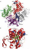
Locations of Pol δ amino acids implicated in segmental forms of progeria. A: The Pol δ structure from S. cerevisiae is rendered as a ribbon diagram [Swan et al., 2009b] (Protein Data Bank accession code 3IAY). Amino acid positions are numbered by the corresponding human position. Progeria-associated amino acids (R507 and S605) are shown as teal CPK spheres. Polymerase domains and other structural elements are color coded: amino domain, gray; exonuclease (exo), red; palm domain, purple; fingers domain, blue; thumb domain, green; primer template DNA, tan sticks; dNTP, orange CPK sticks; catalytic carboxylate residues, green CPK sticks; a disordered loop (486–491) in the exonuclease domain, orange dashed line. The palm and fingers domains together form the polymerase catalytic active site. B: Location of R507C in the Pol δ exonuclease domain compared with cancer-associated amino acid substitutions. Superimposed on the Pol δ exo domain are the last three nucleotides of the primer strand (yellow CPK sticks) and coordinating metal ions (green spheres) from the editing structure of the highly related polymerase, RB69 Pol [Shamoo and Steitz, 1999] (Protein Data Bank accession code 1CLQ). Cancer-associated substitutions are gray CPK spheres. Amino acids that mediate binding of the primer strand or interact with R507 are red CPK spheres. αG and αI are α-helices discussed in the text.
Figure 4
Figure 4
Chromosomal stability in LCLs of POLD1-mutated patients. Shows the mean number of aberrations per cell observed in 100 metaphases of an unaffected individual (AG1010), two individuals bearing a p.605Sdel mutation (COLOM1010 and JAE91301), an individual bearing a p.R507C mutation (MINN1010), and a Werner syndrome patient bearing a mutation in WRN gene (MY1010). LCLs were untreated or treated either with 40 ng/ml of MMC (mitomycin C) or 0.3 μM of APH (aphidicolin). Bar graphs summarize two independent experiments. P values are relative to AG1010 for each treatment (n.s., not significant, ***P < 0.001).

Similar articles

Cited by

References

    1. Agarwal AK, Fryns JP, Auchus RJ, Garg A. Zinc metalloproteinase, ZMPSTE24, is mutated in mandibuloacral dysplasia. Hum Mol Genet. 2003;12:1995–2001. - PubMed
    1. Barrios Sanjuanelo A, Munoz Otero C. Atypical Werner syndrome: atypical progeroid syndrome: a case report. An Pediatr (Barc) 2010;73:94–97. - PubMed
    1. Chen L, Lee L, Kudlow BA, Dos Santos HG, Sletvold O, Shafeghati Y, Botha EG, Garg A, Hanson NB, Martin GM, Mian IS, Kennedy BK, Oshima J. LMNA mutations in atypical Werner’s syndrome. Lancet. 2003;362:440–445. - PubMed
    1. Church DN, Briggs SE, Palles C, Domingo E, Kearsey SJ, Grimes JM, Gorman M, Martin L, Howarth KM, Hodgson SV, The NSECG Collaborators. Kaur K, et al. DNA polymerase epsilon and delta exonuclease domain mutations in endometrial cancer. Hum Mol Genet. 2013;22:2820–2828. - PMC - PubMed
    1. Hisama FM, Lessel D, Leistritz D, Friedrich K, McBride KL, Pastore MT, Gottesman GS, Saha B, Martin GM, Kubisch C, Oshima J. Coronary artery disease in a Werner syndrome-like form of progeria characterized by low levels of progerin, a splice variant of lamin A. Am J Med Genet A. 2011;155A:3002–3006. - PMC - PubMed

Publication types

MeSH terms