. 2015 Jul 14;10(7):e0133152.
doi: 10.1371/journal.pone.0133152. eCollection 2015.

IGF1R- and ROR1-Specific CAR T Cells as a Potential Therapy for High Risk Sarcomas

Affiliations

IGF1R- and ROR1-Specific CAR T Cells as a Potential Therapy for High Risk Sarcomas

Xin Huang et al. PLoS One. .

Abstract

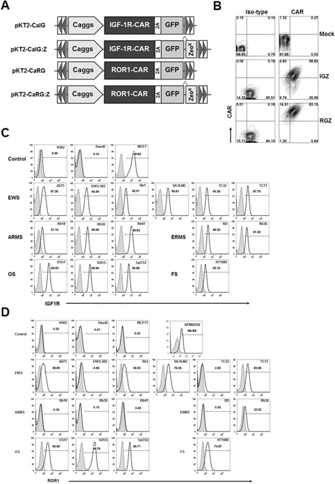
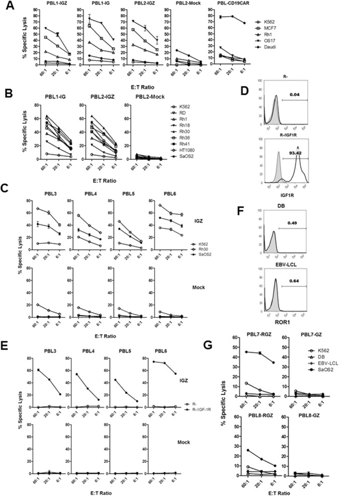
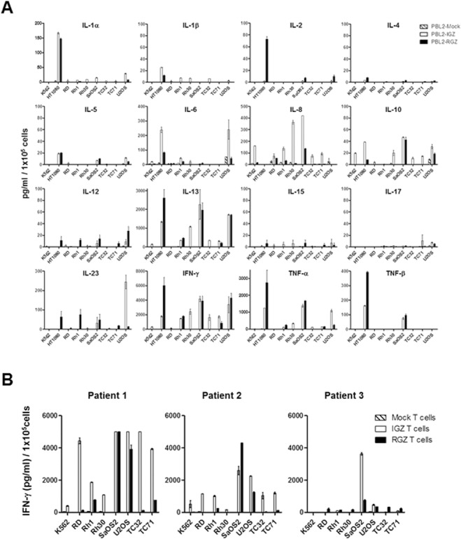
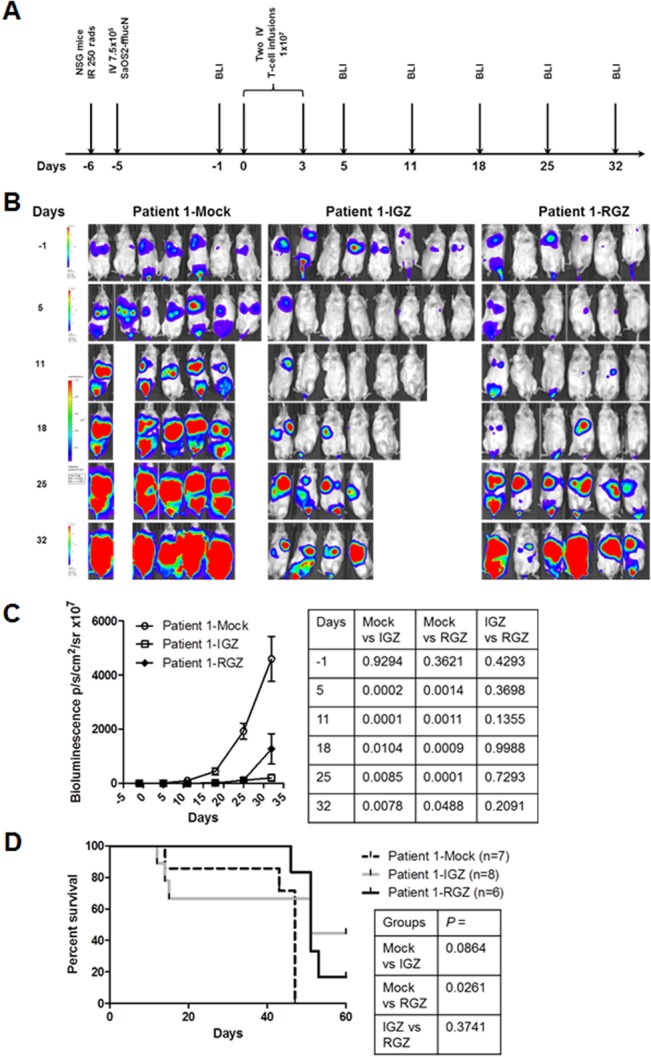
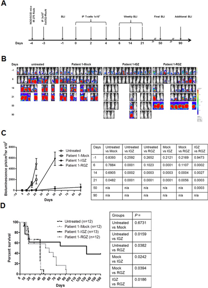
Patients with metastatic or recurrent and refractory sarcomas have a dismal prognosis. Therefore, new targeted therapies are urgently needed. This study was designed to evaluate chimeric antigen receptor (CAR) T cells targeting the type I insulin-like growth factor receptor (IGF1R) or tyrosine kinase-like orphan receptor 1 (ROR1) molecules for their therapeutic potential against sarcomas. Here, we report that IGF1R (15/15) and ROR1 (11/15) were highly expressed in sarcoma cell lines including Ewing sarcoma, osteosarcoma, alveolar or embryonal rhabdomyosarcoma, and fibrosarcoma. IGF1R and ROR1 CAR T cells derived from eight healthy donors using the Sleeping Beauty (SB) transposon system were cytotoxic against sarcoma cells and produced high levels of IFN-γ, TNF-α and IL-13 in an antigen-specific manner. IGF1R and ROR1 CAR T cells generated from three sarcoma patients released significant amounts of IFN-γ in response to sarcoma stimulation. The adoptive transfer of IGF1R and ROR1 CAR T cells derived from a sarcoma patient significantly reduced tumor growth in pre-established, systemically disseminated and localized osteosarcoma xenograft models in NSG mice. Infusion of IGF1R and ROR1 CAR T cells also prolonged animal survival in a localized sarcoma model using NOD/scid mice. Our data indicate that both IGF1R and ROR1 can be effectively targeted by SB modified CAR T cells and that such CAR T cells may be useful in the treatment of high risk sarcoma patients.


Conflict of interest statement

Competing Interests: The authors have declared that no competing interests exist.

Figures

Fig 1
Fig 1. Cell surface expression of chimeric antigen receptor (CAR) and GFP after transfection and expression of IGF1R and ROR1 in a panel of sarcoma cell lines.
(A) Schematic representation of the Sleeping Beauty (SB) transposons encoding IGF1R CAR and GFP (pKT2-CaIG), IGF1R and GFP:Zeocin (pKT2-CaIG:Z), ROR1 CAR and GFP (pKT2-CaRG), and ROR1 CAR and GFP:Zeocin (pKT2-CaRG:Z). The CAR contains the 4-1BB signaling domain (not shown). A Caggs promoter and a “self-cleaving” 2A peptide flanking the CAR sequence were used to regulate co-expression of CARs and GFP or GFP-Zeocin fusion in the SB transposon trans vectors, namely pKT2-CaIG, pKT2-CaRG, pKT2-CaIG:Z, and pKT2-CaRG:Z. (B) Expression of CAR and GFP in T cells derived from a sarcoma patient (Patient 1) after transfection of PBMCs with pKT2-CaIG:Z/pCMV-SB100X or pKT2-CaRG:Z/pCMV-SB100X or no DNA (as mock) and selection with zeocin. Similar data were obtained in T cells from other two patients with sarcomas and two healthy donors (data not shown). (C) Flow cytometric analysis of IGF1R expression in sarcoma cell lines including Ewing sarcoma (EWS), alveolar or embryonal rhabdomyosarcoma (ARMS or ERMS), osteosarcoma (OS), and fibrosarcoma (FS). K562 (erythroleukemia), Daudi (B-cell Burkitt lymphoma), MCF7 (breast cancer) cell lines were used as control. (D) Flow cytometric analysis of ROR1 expression in sarcoma cell lines. RPMI8226 (multiple myeloma) cell line was used as control. Similar data shown in (C) and (D) were obtained in at least three independent assays.
Fig 2
Fig 2. In vitro anti-sarcoma cytotoxicity of IGF1R and ROR1 CAR T cells.
(A) Cytotoxicity against IGF1R+ target cells including sarcoma cell lines by SB modified IGF1R CAR T cells derived from two healthy donors. PBL1-IGZ, PBL1-IG, PBL2-IGZ and PBL2-mock T cells were generated by transfection of PBMCs derived from two healthy donors (PBL1 and PBL2) using pKT2-CaIG:Z or pKT2-CaIG plus pCMV-SB100X plasmids or without DNA. PBL-CD19CAR T cells were used as control. PBL1-IGZ: Rh1 vs K562 and OS17 vs K562, p < 0.001 at all 3 E/T ratios. PBL1-IG: Rh1 vs K562 and OS17 vs K562, p < 0.01. PBL2-IGZ: Rh1 vs K562, p < 0.001, OS17 vs K562, p < 0.01. Killing of Rh1 and OS17 in each CAR T cell group was also significant compared to the corresponding tumor cells in mock T cell group (p < 0.05). (B) Cytotoxicity of IGF1R CAR T cells against a panel of sarcoma cell lines. Comparisons in PBL1-IGZ and PBL2-IGZ were conducted between each sarcoma line and K562, P = 0.0001 at all E/T ratios. (C) Cytotoxicity against sarcoma cells by IGF1R CAR T cells derived from four more healthy donors but not by mock T cells. Comparisons in PBL3-IGZ, PBL4-IGZ, PBL5-IGZ and PBL6-IGZ were conducted between Rh30 vs K562 and SaOS2 vs K562, P = 0.0001 at all E/T ratios.(D) Expression of human IGF1R in R- transfected cell line confirmed by flow cytometry. (E) Specific cytotoxicity against human IGF1R transfected cell line by IGF1R CAR T cells. Comparisons in PBL3-IGZ, PBL4-IGZ, PBL5-IGZ and PBL6-IGZ were conducted between R- and R-/IGF1R, P = 0.0001 at all E/T ratios. (F) Expression of ROR1 in DB and RBV-LCL cell lines. ROR1- K562 and ROR1+ RPMI8226 were used as control (data not shown). (G) Cytotoxicity against ROR1+ target cells including a sarcoma cell line by ROR1 CAR T cells derived from two healthy donors. Comparisons in PBL7-RGZ, PBL7-GZ, PBL8-RGZ and PBL8-GZ were conducted between each target cell and K562 at all E/T ratios. PBL7-RGZ: SaOS2 vs K562, p = 0.0001, 0.0008, 0.0001 (E/T of 60:1, 20:1 and 6:1). PBL7-GZ: SaOS2 vs K562, p = 0.0221, 0.0762, 0.1121. PBL8-RGZ: SaOS2 vs K562, p = 0.0001, 0.0009, 0.0041. PBL8-GZ: SAOS2 vs K562, p = 0.2585, 0.8439, 0.9298. P values for DB vs K562 and EBV-LCL vs K562 were not shown. All cytotoxicity data shown are mean ± S.E. of triplicates.
Fig 3
Fig 3. Cytokine profiling of IGF1R and ROR1 CAR T cells in response to sarcoma stimulation.
(A) Significant production of IFN-γ, TNF-α, and IL-13 by IGF1R and ROR1 CAR T cells derived from a healthy donor. Data shown are mean ± S.E. of duplicates. One out of two representative data is shown. (B) Antigen-specific production of IFN-γ by IGF1R and ROR1 CAR T cells derived from three sarcoma patients. Data shown are mean ± S.E. of duplicates.
Fig 4
Fig 4. In vivo anti-sarcoma activity of IGF1R and ROR1 CAR T cells in a disseminated mouse model.
(A) The experimental schedule of tumor cell injection, CAR T cell infusion and BLI monitoring. Prior to testing, all mice displayed normal healthy status. B) Bioluminescent imaging (BLI) of tumor growth in NSG mice (three groups, n = 6–8 each) treated with a sarcoma patient derived T cells expressing IGF1R CAR (IGZ), ROR1 CAR (RGZ) or mock T cells. SaOS2-fflucN cells were transduced with a lentiviral vector expressing humanized firefly luciferase and truncated nerve growth factor receptor (NGFR). Two mice in the mock group died of tumor progression on day 8. Four mice in IGZ group died of unknown causes on day 8, 9, 16 and 24, probably due to cytokine storms. (C) Bioluminescent intensity of the mice treated with the T cells. All p values were shown in the right panel table and were verified independently. (D) Animal survival after T-cell therapy. All p values were determined using Mantel-Haenszel logrank test and shown in the right panel table. The p values were independently confirmed. Note that p > 0.05 between mock vs IGZ was likely due to a small sample size.
Fig 5
Fig 5. In vivo anti-sarcoma response by IGF1R and ROR1 CAR T cells in a localized tumor mouse model.
(A) The experimental schedule of tumor cell injection, CAR T cell infusion and BLI monitoring. Prior to testing, all mice displayed normal healthy status. (B) Bioluminescent imaging (BLI) of tumor growth in NOD/SCID mice (four groups, n = 12–13 each) treated with a sarcoma patient derived T cells expressing IGF1R CAR (IGZ), ROR1 CAR (RGZ) or mock T cells. One group mice were untreated. Two mice in the untreated group died of tumor progression on day 13 and 20 and the other two mice died of unknown causes on day 12 and 13. Three mice in the mock group died of tumor progression on day 7, 12 and 19 andone died of unknown causes on day 13. Three mice in the IGZ group died of unknown causes on day 10 and two died of unknown courses on day 17 and 18. Two mice in the RZG group died of tumor progression on day 12 and 50 andone died of unknown causes on day 13. (C) Bioluminescent intensity of the mice treated with the T cells. All p values were shown in the right panel table and were verified independently. (D) Animal survival after T-cell therapy. All p values were determined by the Mantel-Haenszel logrank test and are shown in the right panel table.

References

    1. June CH, Riddell SR, Schumacher TN. Adoptive cellular therapy: A race to the finish line. Science translational medicine. 2015;7(280):280ps7 10.1126/scitranslmed.aaa3643 . - DOI - PubMed
    1. Rosenberg SA, Dudley ME. Adoptive cell therapy for the treatment of patients with metastatic melanoma. Current opinion in immunology. 2009;21(2):233–40. 10.1016/j.coi.2009.03.002 - DOI - PMC - PubMed
    1. Maude SL, Frey N, Shaw PA, Aplenc R, Barrett DM, Bunin NJ, et al. Chimeric antigen receptor T cells for sustained remissions in leukemia. The New England journal of medicine. 2014;371(16):1507–17. 10.1056/NEJMoa1407222 - DOI - PMC - PubMed
    1. Porter DL, Levine BL, Kalos M, Bagg A, June CH. Chimeric antigen receptor-modified T cells in chronic lymphoid leukemia. The New England journal of medicine. 2011;365(8):725–33. 10.1056/NEJMoa1103849 - DOI - PMC - PubMed
    1. Brentjens RJ, Davila ML, Riviere I, Park J, Wang X, Cowell LG, et al. CD19-targeted T cells rapidly induce molecular remissions in adults with chemotherapy-refractory acute lymphoblastic leukemia. Science translational medicine. 2013;5(177):177ra38 10.1126/scitranslmed.3005930 - DOI - PMC - PubMed

Publication types

MeSH terms