. 2015 Aug;96(4):203-31.
doi: 10.1111/iep.12135. Epub 2015 Jul 15.

Fell-Muir Lecture: Heparan sulphate and the art of cell regulation: a polymer chain conducts the protein orchestra

Affiliations

Fell-Muir Lecture: Heparan sulphate and the art of cell regulation: a polymer chain conducts the protein orchestra

John Gallagher. Int J Exp Pathol. 2015 Aug.

Abstract

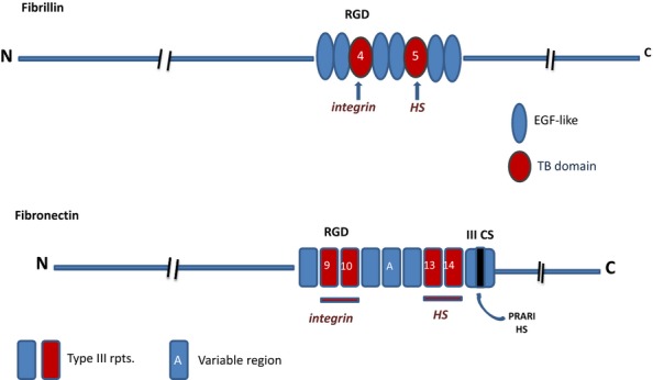
Heparan sulphate (HS) sits at the interface of the cell and the extracellular matrix. It is a member of the glycosaminoglycan family of anionic polysaccharides with unique structural features designed for protein interaction and regulation. Its client proteins include soluble effectors (e.g. growth factors, morphogens, chemokines), membrane receptors and cell adhesion proteins such as fibronectin, fibrillin and various types of collagen. The protein-binding properties of HS, together with its strategic positioning in the pericellular domain, are indicative of key roles in mediating the flow of regulatory signals between cells and their microenvironment. The control of transmembrane signalling is a fundamental element in the complex biology of HS. It seems likely that, in some way, HS orchestrates diverse signalling pathways to facilitate information processing inside the cell. A dictionary definition of an orchestra is 'a large group of musicians who play together on various instruments …' to paraphrase, the HS orchestra is 'a large group of proteins that play together on various receptors'. HS conducts this orchestra to ensure that proteins hit the right notes on their receptors but, in the manner of a true conductor, does it also set 'the musical pulse' and create rhythm and harmony attractive to the cell? This is too big a question to answer but fun to think about as you read this review.

Keywords: glycosaminoglycan; heparan sulphate; heparan sulphate/heparin.


Figures

Figure 1
Figure 1
Cell surface heparan sulphate proteoglycans (HSPGs). The major cell surface HSPGs are the transmembrane syndecans and the GPI-anchored glypicans. The syndecans are constitutive dimers and play key roles in matrix biogenesis, cell adhesion to the extracellular matrix (ECM) and transmission of matrix-derived signals to the cell interior. The glypicans regulate morphogen gradients, signalling and the endocytosis of morphogen receptor complexes; these glypican-related specializations may be facilitated by the close proximity of the heparan sulphate (HS) chains to the cell surface. Both HSPG families are probably involved in binding and activating the many growth factors that utilize an HS co-receptor.
Figure 2
Figure 2
Enzymatic modifications in the biosynthesis of heparan sulphate (HS). The N-acetylated repeat disaccharide unit (a) in the HS precursor, heparan, is converted to HS by a series of modification enzymes (HS-MEs) that act in the following order: NDST, N-deactylase/N-sulphotransferase; C-5 epimerase (converts GlcA to IdoA); 2OST, 2-O-sulphotransferase; 6OST, 6-O-sulphotransferase; and 3OST, 3-O-sulphotransferase. The sequential actions of these enzymes produce a fully modified disaccharide (b) that contains IdoA and sulphate groups at all potential sites of modification. However, the modifications are incomplete at each stage, generally clustered in domains, and give rise to considerable variability in the structure of HS. Extensive regions of the heparan chain remain unmodified. S domains are formed by repeat GlcNS-IdoA, 2S units modified to varying degrees by sulphation at C6 and occasionally at C3. GlcNAc residues may be a target for 6OSTs when positioned next to an N-sulphated unit. As a consequence of this restriction, GlcNAc,6S (c) is found only in (NA)/NS regions of HS.
Figure 3
Figure 3
Domain structure of heparan sulphate (HS). The models illustrate a typical HS species from mammalian cells, rat liver and Drosophila. Mammalian HS is an ordered structure composed of an alternating arrangement of hypervariable sulphated regions [S- and N-acetylated (NA)/NS domains] and non-sulphated regions (NA domains) spaced in a fairly regular manner along the polymer; chain lengths vary from about 50 to 200 disaccharide units. An internal NA domain of approximately 10 disaccharides is contiguous with the glycosaminoglycan–protein linkage sequence. An S domain, often highly sulphated, is common at the distal, non-reducing end of the chain. Rat liver HS is a notable exception to the general design of mammalian HS species; it is an asymmetric structure with three, closely spaced S domains arranged towards the chain periphery but with retention of the internal, non-sulphated NA domain. HS synthesized by Drosophila is a relatively short, two-domain polymer in which a core NA sequence is connected to a longer, heparin-like distal region (Kusche-Gullberg et al. 2012). HS thus appears to have acquired a more complex structure during the course of evolution with an extension of chain length accompanied by the emergence of internal sulphated regions but with retention of the core NA domain.
Figure 4
Figure 4
Molecular model of a flexible N-acetylated (NA) region of heparin sulphate flanked by short S domains. The model representing a long NA region in heparan sulphate (HS) was made on the basis of one of the models in the ensemble 4KHL.pdb, currently available as supplementary material to Khan et al. J. Biol. Chem. 2013, 288:27737–27751. This was a 24-mer of the heparan GlcA-GlcNAc sequence, consistent with X-ray scattering results. The two short S domains added at each end are made up of trisaccharides from the NMR structure of heparin, HPN1.pdb. This representation is an illustration, not the results of a simulation exercise. The model was kindly prepared by Professor Barbara Mulloy.
Figure 5
Figure 5
Crystal structure of a heparin dp6/FGF2 complex (Faham et al. 1996). The model shows the structure of FGF2 and a heparin hexasaccharide from the pdb file 1BFC.pdb. The protein is shown as a solid ribbon coloured by secondary structure: blue for beta-strands, red for helices, green for turns and white otherwise. Water molecules are red circles. FGF, fibroblast growth factors.
Figure 6
Figure 6
The interaction of FGF2 and FGF1 with heparin. (a) Sequence alignment of FGF2 and FGF1 in the main heparin/heparan sulphate binding sequence; conserved residues involved in heparin binding are in bold text. (b) Schematic diagram of the main FGF2 heparin contacts in the co-crystal FGF2 heparin complex in Figure 4. The GlcNS-IdoA,2S sequence (red) interacts with a high affinity subsite in FGF2; R121 are K126 are critical residues in this site. The predominant interactions are electrostatic, but Asn (N28 and N102) and Gln (Q131) participate in important H-bonds with the bound heparin. In the crystal structure, the IdoA,2S residue in the high-affinity site is in 1C4 chair conformation and the non-sulphated IdoA is in the 2SO skew boat conformer. For simplicity, non-interacting 6-O-sulphate groups on the amino sugars are not shown. FGF, fibroblast growth factors.
Figure 7
Figure 7
Sulphation clusters in heparin and HS: FGF1- and FGF2-binding sites. The diagrams follow the proposals of Pellegrini (2001) and Mulloy (2005) for illustrating the disposition of sulphate groups in the heparin helix. In the dp8 fragments, disaccharide repeats of iduronate (ovals) and glucosamine (rectangles) are inverted to show the clusters of three sulphates (NS, 2S, 6S) in sequences of three residues on either side of the molecule. Heparin dp8 is shown as a fully sulphated molecule. The HS dp8 fragments have a lower degree of sulphation than heparin. The proposed minimal binding sites for FGF1 and FGF2 extend over a similar sugar sequence of five monosaccharides but differ in the required degree of sulphation. FGF monomers bind to only one side of the saccharide chain. In the asymmetric model of a proposed mitogenically active configuration of FGF as shown in Figure 8, the growth factor assembles on HS in a trans-dimer arrangement. FGF1 dimers form on HS sequences with two trisulphation clusters as shown. Although monomeric binding of FGF2 is not dependent on 6-sulphates, at least one 6S group is required for activation. This key 6S may be positioned towards the end of a bioactive sequence where it could interact with an FGF2 monomer that binds with opposite polarity to that shown for FGF2 in the primary binding site (see text for details). FGF, fibroblast growth factors; HS, heparan sulphate.
Figure 8
Figure 8
Diagrammatic models of the crystal structures of FGF/FGFR/(D2 and D3 domains)/heparin complexes. In the asymmetric model (Pellegrini et al. 2000), two FGFs bind on opposite sides of a heparin dp10 saccharide and recruit two FGFRs in a stable 2:2:1 complex with minimal protein:protein contacts. In the symmetric model (Schlessinger et al. 2000), two half complexes (1:1:1 FGF:FGFR:dp10) assemble at the non-reducing ends of two dp10 heparin saccharides and these then combine, primarily by means of extensive FGFR interactions, to form the symmetric complex. In the diagrams, the heparin saccharides in the crystal structures are imagined as S domains in HS positioned internally in the asymmetric model or at the periphery of the HS chain in the symmetric version. FGF, fibroblast growth factors; HS, heparan sulphate.
Figure 9
Figure 9
Hepatocyte growth factor/scatter factor (HGF/SF), its splice variants and interactions with heparan sulphate (HS). (a) HGF/SF is a disulphide-linked heterodimer with an N-terminal hairpin loop (N), four kringle domains (K1–K4) and an inactive serine protease (SP) domain. The primary HS-binding site is in the hairpin loop, with accessory sites in the K1 and SP regions. NK1 and NK2 are splice variants of the hgf/sf gene. (b) HS S domains of length dp12-dp14 are the optimum size for high-affinity binding to HGF/SF. In principle, HS fragments of this length are sufficient to engage in a three-point attachment to HGF/SF that may stabilize an active conformation of the modular elements in the native protein. (c) NK1 has an absolute requirement for HS or heparin to bind the Met receptor and for signalling activity in cultured cells. In crystal structures, NK1 forms dimers in the presence of heparin and four sulphated monosaccharides make contact with the binding site in the N-domain. Heparin (or HS) may stabilize the dimer and/or expose the dimerization surfaces in the N and K1 regions. The tendency for NK1 domains to form stable interactions in the presence of heparin suggests a mechanism for dimerization and activation of native HGF/SF.
Figure 10
Figure 10
Heparan sulphate-binding sites in the midkine (MK) dimer. In the head-to-head midkine dimer, two CW motifs (+++) form an extended heparan sulphate (HS)-binding site at the dimer interface; in each monomer, the CW motif slopes towards three additional basic residues that further enhance HS affinity. Cell surface HS stabilizes the midkine dimer and is essential for midkine signalling.
Figure 11
Figure 11
Heparin-/heparan sulphate (HS)-binding sites in the VEGF165 heparin-binding domain (HBD) and in the VEGF165 dimer. (a) Ribbon diagram of the VEGF165 HBD (residues 111–165; Protein Data Bank code 2VGH) with a docked heparin dp7 [space-filling representation: carbon (grey), oxygen (red), sulphur (yellow), nitrogen (blue) and hydrogen (white)]. Basic residues lining the shallow binding groove are shown in a stick representation (green). (b) The same complex with 2VGH depicted as a protein surface. Arg and Lys residues are shown in blue, and Glu and Asp residues are shown in red. The heparin dp7 saccharide is a stick representation. The atomic coloration is as in (a), except that carbons are shown in green. (c) A K5 lyase-resistant HS fragment (white and hatched boxes) is bound to the HBDs of VEGF165 homodimer (grey). The N- and C-termini of VEGF165 and the reducing (R) and nonreducing (NR) ends of the HS chain are indicated. The VEGF165 subunits are held by disulfide bonds in an antiparallel ‘side-by-side’ orientation. Arrows indicate plasmin cleavage at the sites that release the HBDs. Basic residues in each heparin-binding cleft are shown (+). The two clefts are occupied by separate S domains (white boxes) in the same HS chain. The S domains are at least dp6 in length and are 6-O-sulphated. This research was originally published in Journal of Biological Chemistry. Robinson et al. © the American Society for Biochemistry and Molecular Biology.” VEGF, vascular endothelial growth factor.
Figure 12
Figure 12
Models of CXCL chemokines in complexes with heparan sulphate (HS) and heparin. The models proposed for HS in complexes with PF4 and IL-8 are based on the structures of chemokine-binding domains in HS protected from degradation by heparinase enzymes (see text for details). In PF4, the S domains run perpendicular to the alpha-helices but adopt a parallel orientation to the alpha-helices in IL-8. The alpha-helices in SDF1-α are not involved in HS–heparin binding. Molecular docking reveals that heparin (dp12) binds along a positively charged ‘crevasse'at the interface of the SDF1-a dimer and then extends to the N-terminal lysines in each monomer (Sadir et al. 2001). Ref. PF4 model in (a): This research was originally published in Journal of Biological Chemistry. Authors: Sally E. Stringer and John T. Gallagher Title: Specific Binding of the Chemokine Platelet Factor 4 to Heparan Sulfate. J. Biol. Chem. (1997) 272, 20508–20514 © the American Society for Biochemistry and Molecular Biology.”
Figure 13
Figure 13
Cell- and HS-binding regions of fibrillin and fibronectin. The diagram illustrates the similarity in arrangement of the major integrin and HS-binding regions of fibrillin and fibronectin. The co-operative interactions of these sites with cell surface integrins and HSPGs are essential for cell attachment to the ECM and for matrix-driven focal adhesions and signalling.
Figure 14
Figure 14
(a) Heparan sulphate (HS)/heparin binding sites in the N-terminal domain (NTD) and helical region of the α1 chain of collagen XI. Heparan sulphate-/heparin-binding regions in collagen α1(XI)-chain are found in the 223-residue globular Npp domain in the form of an XBBBXXBX CW motif, in the highly basic variable region, and in two sites in the major triple helix including a similar sequence to the 905–921 residue sequence present in the collagen type α1(V)-chain. The CW motif in the Npp domain is also present in collagen α1(V). The NTD of the α1-chain is retained in the collagen XI heterotrimeric triple helix and projects from the surface of the collagen fibril. The NTDs of the α2- and α3-chains are rapidly removed by proteolysis before assembly of the collagen XI monomer into fibrils. (b) Schematic diagram of the binding sites in synaptic collagen Q. Collagen Q is found only in the neuro-muscular synapse. It contains two (XBBXBX) CW motifs in the triple helix that interact with perlecan HS in the synaptic cleft. The non-collagenous C-terminal region binds to the muscle-specific Musk receptor. The three non-helical N-terminal regions bind four AchE subunits in an asymmetric A12/Q complex that degrades Ach and controls the strength and duration of synaptic transmission.

References

    1. Ai X, Do AT, Lozynska O, Kusche-Gullberg M, Lindahl U. Emerson CP. Qsulf1 remodels the 6-O sulfation states of cell surface heparan sulfate proteoglycans to promote Wnt signaling. J. Cell Biol. 2003;162:341–351. - PMC - PubMed
    1. Ai X, Kitazawa T, Do AT, Kusche-Gullberg M, Labosky PA. Emerson CP., Jr SULF1 and SULF2 regulate heparan sulfate-mediated GDNF signaling for esophageal innervation. Development. 2007;134:3327–3338. - PubMed
    1. Alfano I, Vora P, Mummery R, Mulloy B. Rider C. The major determinant of the heparin binding of glial cell-line-derived neurotrophic factor is near the N-terminus and is dispensable for receptor binding. Biochem. J. 2007;404:131–140. - PMC - PubMed
    1. Allen BL. Rapraeger AC. Spatial and temporal expression of heparan sulfate in mouse development regulates FGF and FGF receptor assembly. J. Cell Biol. 2003;163:637–648. - PMC - PubMed
    1. Allen BL, Filla MS. Rapraeger AC. Role of heparan sulfate as a tissue-specific regulator of FGF-4 and FGF receptor recognition. J. Cell Biol. 2003;155:845–858. - PMC - PubMed

Publication types

Substances