. 2015 Jul 15;16(1):86.
doi: 10.1186/s12931-015-0245-z.

Evidence for a novel Kit adhesion domain mediating human mast cell adhesion to structural airway cells

Affiliations

Evidence for a novel Kit adhesion domain mediating human mast cell adhesion to structural airway cells

Kevin C Gough et al. Respir Res. .

Abstract

Background: Human lung mast cells (HLMCs) infiltrate the airway epithelium and airway smooth muscle (ASM) in asthmatic airways. The mechanism of HLMC adhesion to both cell types is only partly defined, and adhesion is not inhibited by function-blocking anti-Kit and anti-stem cell factor (SCF) antibodies. Our aim was to identify adhesion molecules expressed by human mast cells that mediate adhesion to human ASM cells (HASMCs) and human airway epithelial cells.

Methods: We used phage-display to isolate single chain Fv (scFv) antibodies with adhesion-blocking properties from rabbits immunised with HLMC and HMC-1 membrane proteins.

Results: Post-immune rabbit serum labelled HLMCs in flow cytometry and inhibited their adhesion to human BEAS-2B epithelial cells. Mast cell-specific scFvs were identified which labelled mast cells but not Jurkat cells by flow cytometry. Of these, one scFv (A1) consistently inhibited mast cell adhesion to HASMCs and BEAS-2B epithelial cells by about 30 %. A1 immunoprecipitated Kit (CD117) from HMC-1 lysates and bound to a human Kit-expressing mouse mast cell line, but did not interfere with SCF-dependent Kit signalling.

Conclusion: Kit contributes to human mast cell adhesion to human airway epithelial cells and HASMCs, but may utilise a previously unidentified adhesion domain that lies outside the SCF binding site. Targeting this adhesion pathway might offer a novel approach for the inhibition of mast cell interactions with structural airway cells, without detrimental effects on Kit signalling in other tissues.


Figures

Fig. 1
Fig. 1
Binding of post immune rabbit sera to HLMC. Flow cytometry showing that the post immune serum from a rabbit immunised with HLMC/HMC-1 plasma membranes (bottom row) demonstrates increased binding to HLMC compared to the pre-immune serum (top row) at equivalent dilutions. Representative of data from two immunised rabbits
Fig. 2
Fig. 2
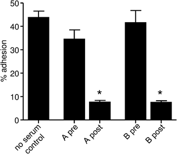
Inhibition of HLMC adhesion to BEAS-2B by post immune rabbit sera. Adhesion of HLMC to BEAS-2B human bronchial epithelial cells was attenuated by the post immune serum from two rabbits (A and B) immunised with HLMC/HMC-1 plasma membranes. n = 5 HLMC donors studied in 5 independent experiments. *p = 0.004 for each
Fig. 3
Fig. 3
The isolation of scFvs that bind to mast cells. a Binding of polyclonal phage-displayed antibodies to HMC-1. Three rounds of panning were carried out, with each round including a negative selection of phage against epitope-masked HMC-1 cells, followed by a positive selection against unmasked HMC-1. HMC-1 cells were detected with 1010 phage from panning rounds 0, 1, 2, and 3 (R0 to R3). Bound scFv was detected with biotin labelled anti-fd antibody and then extravadin-AP conjugate. Absorbances were measured at 405 nm. Individual scFvs that bound to HMC-1 cells were selected by phage ELISA. b and c scFv binding was confirmed by flow cytometry. HMC-1 or Jurkat cells were incubated with purified scFv and binding was visualised with anti-C-myc 9E10 antibody and anti-mouse antibody-FITC conjugate. The mean fluorescence produced with each scFv is expressed as a fold-increase from that produced in the absence of scFv b. ScFvs A1, A3, A8, B1, C1 and D1 were selected against HMC-1; the scFv E4 is a control scFv which does not bind to HMC-1. Representative examples of flow cytometry histograms for the binding of scFvs to HMC-1 and Jurkat cells are shown for antibody A1 and for a no scFv control c. The binding of scFvs to HMC-1 was repeated a minimum of 4 times and all 6 scFvs demonstrated clear binding to HMC-1 cells on each occasion. d scFv A1 also bound to HLMCs when compared to E4 (representative of 3 independent experiments using 3 HLMC donors)
Fig. 4
Fig. 4
The effects of scFv A1 on mast cell adhesion. a HMC-1 adhesion to monolayers of BEAS-2B human airway epithelial cells and primary HASMCs was assessed in the presence of anti-mast cell scFv A1 or a control scFv E4 which does not bind. Baseline adhesion and adhesion in control media containing antibody vehicle (glycerol, final concentration 0.8-1.5 % depending on the scFv batch) is also shown. Plotted as mean ± SEM for all experiments; n = 8 ASM donors used in independent experiments and n = 4 independent experiments for BEAS-2B. *p = 0.048 compared to E4, **p = 0.008 compared to E4. b HLMC adhesion to monolayers of BEAS-2B human airway epithelial cells and primary HASMCs was assessed in the presence of anti-mast cell scFv A1 or a control scFv E4 which does not bind. Baseline adhesion and adhesion in control media containing antibody vehicle (glycerol, final concentration 0.8-1.5 % depending on the scFv batch) is also shown for HASMCs, baseline adhesion is shown for BEAS-2B cells (glycerol not assessed). Plotted as mean ± SEM for all experiments; n = 1 ASM donor and 7 HLMC donors used in n = 7 independent experiments, and n = 6 HLMC donors in independent experiments for BEAS-2B. *p = 0.029 compared to E4, # p = 0.017 compared to E4
Fig. 5
Fig. 5
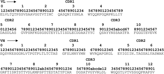
Deduced amino acid sequence of the anti-mast cell scFv A1. The alignment is according to the method of Kabat et al. [18]
Fig. 6
Fig. 6
Identification of Kit as the antigen for scFv A1. a Immunoprecipitation of HMC-1 plasma membrane proteins with scFv A1 identifed a unique band of approximately 145 kDa (arrow). Analogous immunoprecipitations using scFvs C1 (anti-mast cell scFv) and E4 (scFv with irrelevant binding) are shown for comparison. Excision and sequencing demonstrated that this was Kit. b Western blotting of protein immunoprecipitated by scFv A1 demonstrated positive staining using an anti-Kit antibody, with a doublet of the expected size for Kit (145/120 kDa). c Flow cytometry histograms demonstrating that following incubation of HMC-1 with SCF (100 ng/ml) for 15 min, binding of scFv A1 was markedly reduced compared to control, in keeping with SCF-induced Kit internalisation
Fig. 7
Fig. 7
scFv1 A1 binds to human Kit-expressing MCBS1 mouse mast cells. Kit expression was confirmed by flow cytometry (a) and immunofluorescence (b). a Fluorescent histograms (red lines), plotted with corresponding isotype controls (blue line) demonstrate A1 and Kit expression in the W1-AA677 human Kit-expressing mouse mast cell line, but not in E1-AA685 mock-transfected cells or non-transfected control cells. The mean fluorescent intensity was determined minus the total binding of matched isotype controls for A1 and Kit, scFv E4 and mouse IgG respectively. Representative of 3 independent experiments. b Immunofluorescent staining of W1-AA677, E1-AA685 and control cells. Nuclei stained blue with DAPI, A1 and Kit stained red (RPE); inset: isotype control. Representative of two independent experiments
Fig. 8
Fig. 8
scFv A1 does not initiate or interfere with Kit signalling. HMC-1 were incubated with scFv A1 or E4 control in the presence or absence of SCF 100 ng/ml. Total intracellular phosphorylation assessed by Western blotting (PY99 antibody) was not altered by A1 in either the absence or presence of SCF. Phosphorylation of Kit (bands 145/120 kDa) in the left panel was also not altered. Staining for Kit (E1, right panel) and β-actin demonstrates equal loading for Kit within each pair of experiments. Representative of three independent experiments

Similar articles

Cited by

References

    1. Bradding P, Walls AF, Holgate ST. The role of the mast cell in the pathophysiology of asthma. J Allergy Clin Immunol. 2006;117:1277–84. doi: 10.1016/j.jaci.2006.02.039. - DOI - PubMed
    1. Brightling CE, Bradding P, Symon FA, Holgate ST, Wardlaw AJ, Pavord ID. Mast cell infiltration of airway smooth muscle in asthma. N Engl J Med. 2002;346:1699–705. doi: 10.1056/NEJMoa012705. - DOI - PubMed
    1. Bradding P, Roberts JA, Britten KM, Montefort S, Djukanovic R, Mueller R, Heusser CH, Howarth PH, Holgate ST. Interleukin-4, −5, and −6 and tumor necrosis factor-alpha in normal and asthmatic airways: evidence for the human mast cell as a source of these cytokines. Am J Respir Cell Mol Biol. 1994;10:471–80. doi: 10.1165/ajrcmb.10.5.8179909. - DOI - PubMed
    1. Carroll NG, Mutavdzic S, James AL. Increased mast cells and neutrophils in submucosal mucous glands and mucus plugging in patients with asthma. Thorax. 2002;57:677–82. doi: 10.1136/thorax.57.8.677. - DOI - PMC - PubMed
    1. Yang W, Kaur D, Okayama Y, Ito A, Wardlaw AJ, Brightling CE, Bradding P. Human lung mast cells adhere to human airway smooth muscle, in part, via tumor suppressor in lung cancer-1. J Immunol. 2006;176:1238–43. doi: 10.4049/jimmunol.176.2.1238. - DOI - PubMed

Publication types

Substances

LinkOut - more resources