. 2015 Jul 14;6(4):e00705.
doi: 10.1128/mBio.00705-15.

The Endoplasmic Reticulum Stress Sensor Inositol-Requiring Enzyme 1α Augments Bacterial Killing through Sustained Oxidant Production

Affiliations

The Endoplasmic Reticulum Stress Sensor Inositol-Requiring Enzyme 1α Augments Bacterial Killing through Sustained Oxidant Production

Basel H Abuaita et al. mBio. .

Abstract

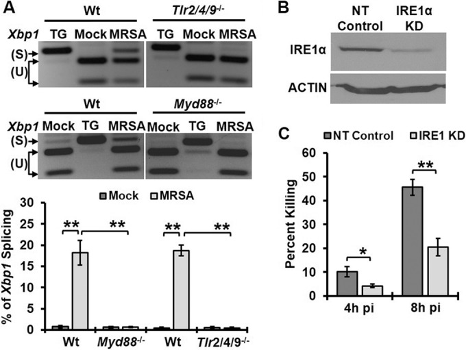
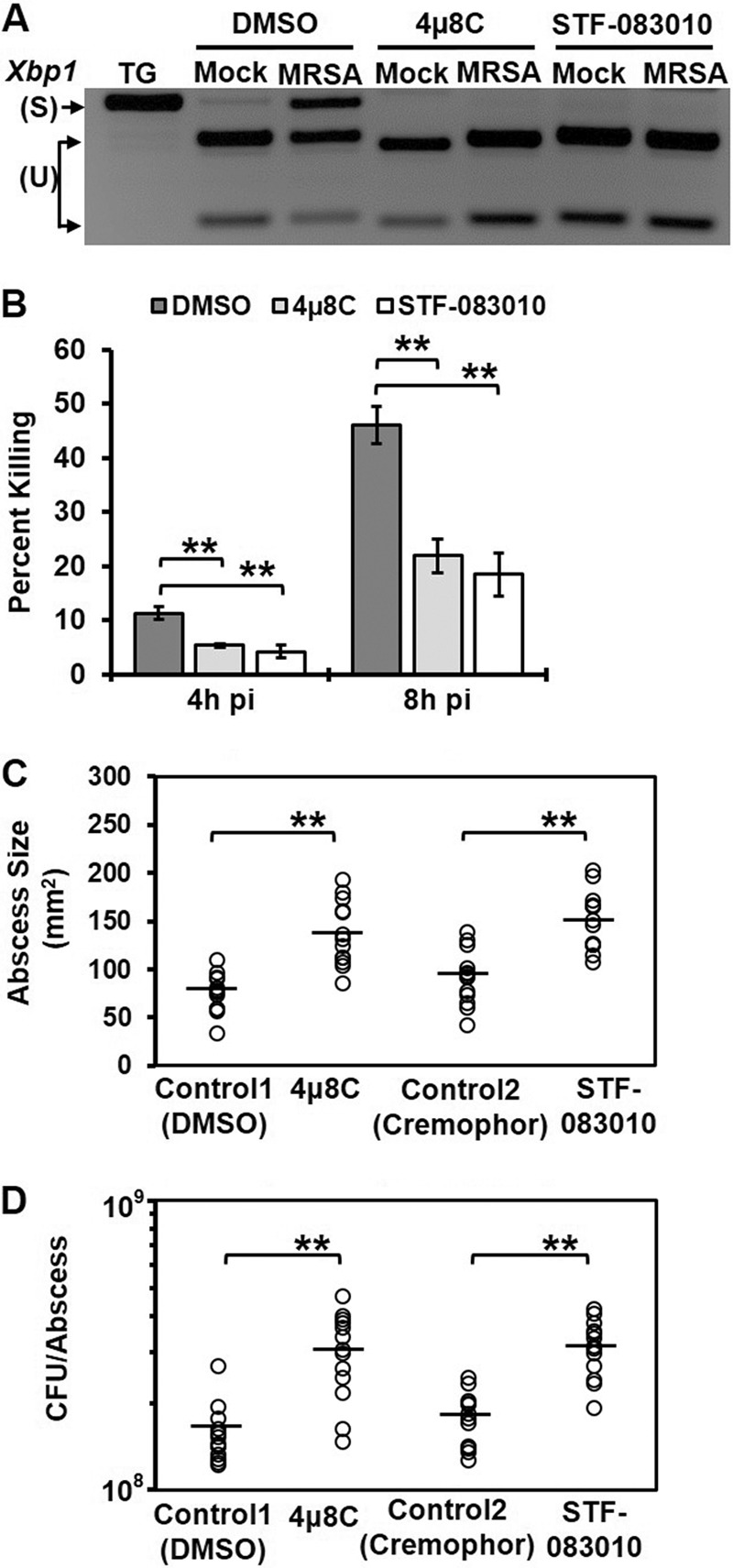
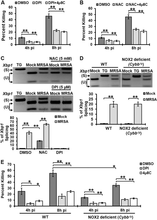
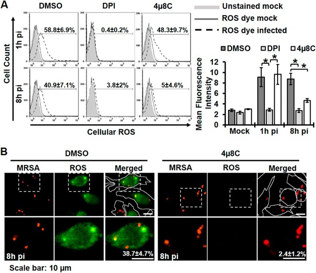
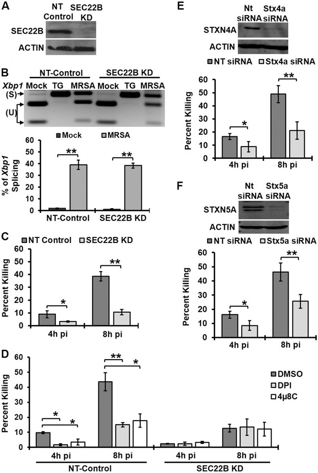
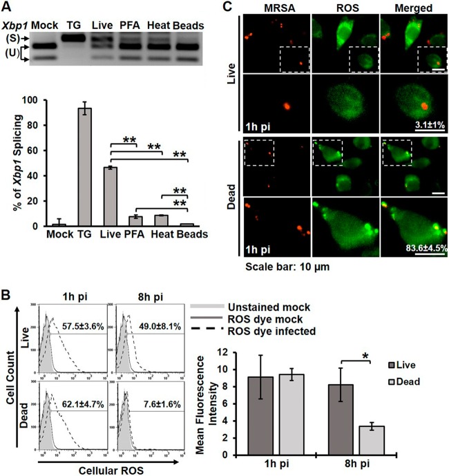
Bacterial infection can trigger cellular stress programs, such as the unfolded protein response (UPR), which occurs when misfolded proteins accumulate within the endoplasmic reticulum (ER). Here, we used the human pathogen methicillin-resistant Staphylococcus aureus (MRSA) as an infection model to probe how ER stress promotes antimicrobial function. MRSA infection activated the most highly conserved unfolded protein response sensor, inositol-requiring enzyme 1α (IRE1α), which was necessary for robust bacterial killing in vitro and in vivo. The macrophage IRE1-dependent bactericidal activity required reactive oxygen species (ROS). Viable MRSA cells excluded ROS from the nascent phagosome and strongly triggered IRE1 activation, leading to sustained generation of ROS that were largely Nox2 independent. In contrast, dead MRSA showed early colocalization with ROS but was a poor activator of IRE1 and did not trigger sustained ROS generation. The global ROS stimulated by IRE1 signaling was necessary, but not sufficient, for MRSA killing, which also required the ER resident SNARE Sec22B for accumulation of ROS in the phagosomal compartment. Taken together, these results suggest that IRE1-mediated persistent ROS generation might act as a fail-safe mechanism to kill bacterial pathogens that evade the initial macrophage oxidative burst.

Importance: Cellular stress programs have been implicated as important components of the innate immune response to infection. The role of the IRE1 pathway of the ER stress response in immune secretory functions, such as antibody production, is well established, but its contribution to innate immunity is less well defined. Here, we show that infection of macrophages with viable MRSA induces IRE1 activation, leading to bacterial killing. IRE1-dependent bactericidal activity required generation of reactive oxygen species in a sustained manner over hours of infection. The SNARE protein Sec22B, which was previously demonstrated to control ER-phagosome trafficking, was dispensable for IRE1-driven global ROS production but necessary for late ROS accumulation in bacteria-containing phagosomes. Our study highlights a key role for IRE1 in promoting macrophage bactericidal capacity and reveals a fail-safe mechanism that leads to the concentration of antimicrobial effector molecules in the macrophage phagosome.


Figures

FIG 1
FIG 1
IRE1 activated through TLR signaling enhances macrophage bactericidal activity. (A) RT-PCR analysis of Xbp1 mRNA splicing in BMDM when left untreated (mock), treated with 5 µM thapsigargin (TG), or infected with MRSA for 8 h at an MOI of 20. PCR products were digested with PstI endonuclease. Because unspliced mRNA contains a PstI site within the 26 spliced region, the digested PCR products yield two smaller fragments representing unspliced (U) Xbp1 and one larger fragment representing spliced (S) Xbp1. RT-PCR images are representative of ≥3 independent experiments, and the percent spliced Xbp1 was calculated based on band densitometry as follows: [Xbp1s/(Xbp1s + Xbp1u)]. (B) Immunoblots of cell lysate from RAW 264.7 macrophages stably transduced with lentivirus-encoded shRNA for nontarget (NT-control) or IRE1 KD, probed with an anti-IRE1 antibody or anti-actin antibody as a loading control. (C) NT-control and IRE1 KD macrophages were infected with MRSA (MOI, 20). The percent killing was quantified by the percent difference in CFU at the indicated time points relative to results at 2h pi: [(CFU2h pi − CFU4h pi)/CFU2h pi]. Results presented are averages of ≥3 independent experiments ± standard deviations. *, P < 0.05; **, P < 0.01.
FIG 2
FIG 2
IRE1 endonuclease is required for macrophage bactericidal activity and resistance to MRSA infection. (A) RT-PCR analysis results of Xbp1 mRNA splicing in mock- or MRSA-infected RAW 264.7 cells treated with DMSO control or small-molecule IRE1 inhibitors 4µ8C (25 µM) or STF-083010 (60 µM). (B) The percent killing was quantified as described for Fig. 1C and is presented as the average of ≥3 independent experiments ± the standard deviation. RAW 264.7 macrophages were infected with MRSA (MOI, 20) in the presence of the DMSO control or IRE1 inhibitor 4µ8C (25 µM) or STF-083010 (60 µM). (C) Skin abscess size (in square millimeters) from C57BL/6 mice infected subcutaneously with 108 CFU MRSA at 3 days pi. Mice were injected intraperitoneally with DMSO control (5%), 4µ8C (10 mg/kg), cremophor EL control (15%), or STF-083010 (30 mg/kg) 1 day prior to and each day during infection. (D) Bacterial burden in skin abscesses from mice infected as described for panel C. Abscess size and bacterial load are shown for mice infected in 2 independent experiments; horizontal lines represent the means. *, P < 0.05; **, P < 0.01.
FIG 3
FIG 3
IRE1-dependent killing of MRSA occurs through ROS production. (A) Percent MRSA killing by RAW 264.7 macrophages treated with DMSO, 5 µM DPI, or 5 µM DPI plus 25 µM 4µ8C. (B) The percent MRSA killing by RAW 264.7 macrophages treated with DMSO, 5 mM NAC, or 5 mM NAC plus 25 µM 4µ8C. (C) RT-PCR analysis results for Xbp1 mRNA splicing in mock- or MRSA-infected RAW 264.7 macrophages treated with DMSO control, 5 µM DPI, or 5 mM NAC. TG was used as a positive control for IRE1 activation. (D) BMDM isolated from wild-type and Nox2-deficient mice (Cybb−/y) were infected with MRSA (MOI, 20), and spliced Xbp1 was assessed at 8h pi by RT-PCR. TG was used as a positive control for IRE1 activation. (E) Percent MRSA killing by wild-type or Nox2-deficient (Cybb−/y) macrophages in the presence of DMSO, 5 µM DPI, or 25 µM 4µ8C. Graphs illustrate mean results from ≥3 independent experiments with standard deviations. *, P < 0.05; **, P < 0.01.
FIG 4
FIG 4
IRE1 contributes to sustained ROS production by macrophages. (A) Global ROS production as assessed by flow cytometry using CM-H2DCFDA dye. RAW 264.7 macrophages were infected with MRSA (MOI, 20) in the presence of DMSO, 5 µM DPI, or 25 µM 4µ8C. Flow cytometry was performed on live cells treated with CM-H2DCFDA for 45 min prior to analysis. (Left) The percentage of ROS+ cells was determined by gating against stained mock-infected control cells. Representative plots are shown with the mean percentages of ROS+ cells from ≥3 independent experiments ± the standard deviations. (Right) Quantification of mean fluorescence intensity under the indicated conditions was calculated as the geometric mean, using FlowJo software for results from ≥3 independent experiments ± the standard deviation. (B) Cells with ROS+ phagosomes were imaged with an Olympus IX70 inverted live-cell fluorescence microscope. RAW 264.7 macrophages were infected with MRSA-mCherry (MOI, 20) in the presence of DMSO or 25 µM 4µ8C. CM-H2DCFDA was added at 7h pi for 30 min, and cells were imaged at 8h pi. Cells with ROS+ phagosomes were measured by counting cells with at least one ROS-enriched area colocalized with MRSA from at least 100 ROS+ infected cells. Representative images are shown with the mean percentage of cells with ROS+ phagosomes from ≥3 independent experiments ± the standard deviations. Cells are outlined with a white line in the low-magnification merged images for clarification. *, P < 0.05; **, P < 0.01.
FIG 5
FIG 5
Sec22B enhances MRSA killing by macrophages via an IRE1- and ROS-mediated mechanism. (A) Sec22B and actin immunoblots of cell lysates from NT-control and Sec22B KD macrophages. (B) RT-PCR results from an Xbp1 splicing assay of mock-infected and infected NT-control or Sec22B KD macrophages at 8h pi. (C) NT-control and Sec22B macrophages were infected with MRSA (MOI, 20), and the percent killing was quantified. (D) Percentage of MRSA killing by NT-control and Sec22B KD macrophages in the presence of DMSO, 5 µM DPI, or 25 µM 4µ8C. (E) RAW 264.7 cells were transiently transfected with NT-control or Stx4a siRNA. STX4A, and actin immunoblot assays were performed on cell lysates from a representative experiment (top panel).The percent MRSA killing by these transfected cells averaged over 3 independent experiments is shown in the bottom panel. (F) STX5A and actin immunoblotting results with cell lysates (top) and the percent MRSA killing (bottom) from transiently transfected RAW 264.7 cells with NT-control or Stx5a siRNA. The graphs represent mean results from ≥3 independent experiments ± standard deviuations. *, P < 0.05; **, P < 0.01.
FIG 6
FIG 6
Sec 22B controls sustained ROS accumulation in phagosomes. (A) Flow cytometry analysis results for global ROS production in NT-control- and Sec22B KD-infected macrophages. (Left) Representative histogram plots are shown, with the percentage of ROS+ cells. (Right) Geometric mean fluorescence intensity of ROS production. (B) Live cell fluorescent images of NT-control and Sec22B KD macrophages infected with MRSA-mCherry (MOI, 20) and stained with a ROS fluorescence indicator at 8h pi. (C) The percentage of cells with ROS+ phagosomes was quantified from NT-control and Sec22B KD macrophages. The percentage of cells was determined from the number of cells with at least one enriched area of ROS colocalized with MRSA from at least 100 ROS+ infected cells. (D) The percentage of cells with ROS+ phagosomes was quantified from NT-control, STX4A KD, or STX5A KD macrophages as described for panel C. Graphs represent mean results from ≥3 independent experiments ± standard deviations. *, P < 0.05; **, P < 0.01.
FIG 7
FIG 7
Viable MRSA induces stronger IRE1 activation and is required for sustained ROS production. (A) Xbp1 splicing assay of RNA isolated from RAW 264.7 macrophages that were untreated (mock) or treated with TG and infected with live MRSA, stimulated with PFA-fixed MRSA, stimulated with heat-killed MRSA, or stimulated with latex beads. A representative RT-PCR image (top) and the percentage of Xbp1 splicing (bottom) are shown. The graph represents mean results from ≥3 independent experiments ± standard deviations. (B) Flow cytometry histograms of global ROS production by RAW 264.7 macrophages infected with live MRSA or stimulated with dead MRSA. The percentage of ROS+ cells and the mean fluorescent intensity shown represent means of ≥3 independent experiments ± standard deviations. The mean fluorescent intensity is reported as the geometric mean. (C) ROS localization in RAW 264.7 macrophages infected with live MRSA or stimulated with dead MRSA for 1 h. Representative fluorescence images and the percentages of cells with at least one ROS+ phagosome are shown from ≥3 independent experiments ± standard deviations. *, P < 0.05; **, P < 0.01.

Similar articles

Cited by

References

    1. Ron D, Walter P. 2007. Signal integration in the endoplasmic reticulum unfolded protein response. Nat Rev Mol Cell Biol 8:519–529. doi:10.1038/nrm2199. - DOI - PubMed
    1. Korennykh AV, Egea PF, Korostelev AA, Finer-Moore J, Zhang C, Shokat KM, Stroud RM, Walter P. 2009. The unfolded protein response signals through high-order assembly of Ire1. Nature 457:687–693. doi:10.1038/nature07661. - DOI - PMC - PubMed
    1. Calfon M, Zeng H, Urano F, Till JH, Hubbard SR, Harding HP, Clark SG, Ron D. 2002. IRE1 couples endoplasmic reticulum load to secretory capacity by processing the XBP-1 mRNA. Nature 415:92–96. doi:10.1038/415092a. - DOI - PubMed
    1. Lee K, Tirasophon W, Shen X, Michalak M, Prywes R, Okada T, Yoshida H, Mori K, Kaufman RJ. 2002. IRE1-mediated unconventional mRNA splicing and S2P-mediated ATF6 cleavage merge to regulate XBP1 in signaling the unfolded protein response. Genes Dev 16:452–466. doi:10.1101/gad.964702. - DOI - PMC - PubMed
    1. Reimold AM, Iwakoshi NN, Manis J, Vallabhajosyula P, Szomolanyi-Tsuda E, Gravallese EM, Friend D, Grusby MJ, Alt F, Glimcher LH. 2001. Plasma cell differentiation requires the transcription factor XBP-1. Nature 412:300–307. doi:10.1038/35085509. - DOI - PubMed

Publication types

MeSH terms