Review
. 2015 Oct;6(5):366-76.
doi: 10.1017/S2040174415001300. Epub 2015 Jul 15.

The programming of cardiovascular disease

Affiliations
Review

The programming of cardiovascular disease

K L Thornburg. J Dev Orig Health Dis. 2015 Oct.

Abstract

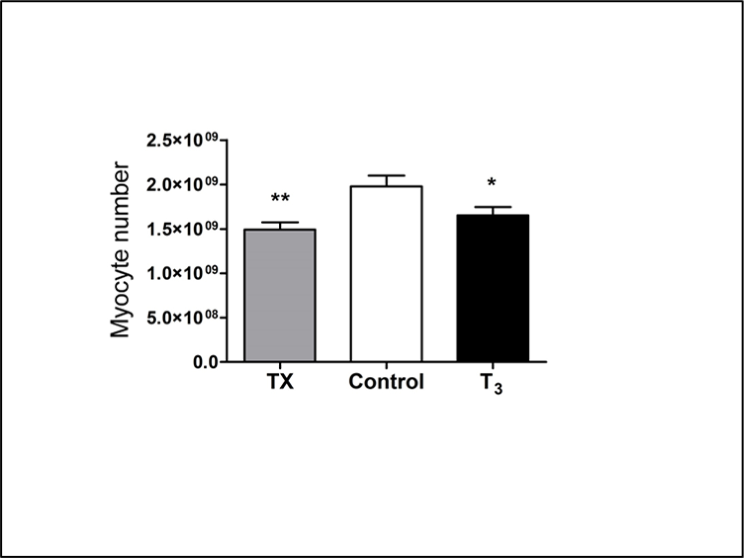
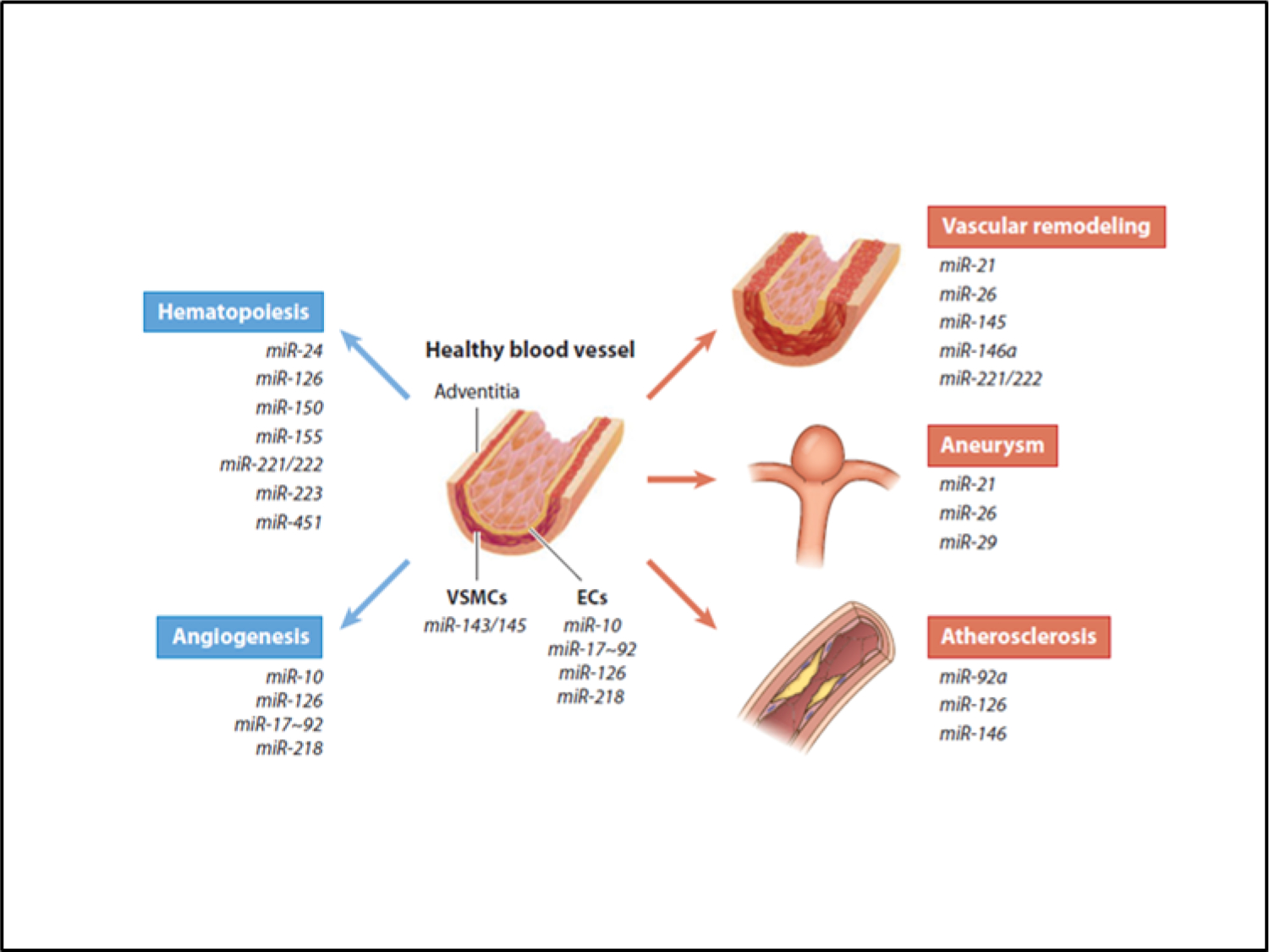
In spite of improving life expectancy over the course of the previous century, the health of the U.S. population is now worsening. Recent increasing rates of type 2 diabetes, obesity and uncontrolled high blood pressure predict a growing incidence of cardiovascular disease and shortened average lifespan. The daily >$1billion current price tag for cardiovascular disease in the United States is expected to double within the next decade or two. Other countries are seeing similar trends. Current popular explanations for these trends are inadequate. Rather, increasingly poor diets in young people and in women during pregnancy are a likely cause of declining health in the U.S. population through a process known as programming. The fetal cardiovascular system is sensitive to poor maternal nutritional conditions during the periconceptional period, in the womb and in early postnatal life. Developmental plasticity accommodates changes in organ systems that lead to endothelial dysfunction, small coronary arteries, stiffer vascular tree, fewer nephrons, fewer cardiomyocytes, coagulopathies and atherogenic blood lipid profiles in fetuses born at the extremes of birthweight. Of equal importance are epigenetic modifications to genes driving important growth regulatory processes. Changes in microRNA, DNA methylation patterns and histone structure have all been implicated in the cardiovascular disease vulnerabilities that cross-generations. Recent experiments offer hope that detrimental epigenetic changes can be prevented or reversed. The large number of studies that provide the foundational concepts for the developmental origins of disease can be traced to the brilliant discoveries of David J.P. Barker.

Keywords: epigenetics; fetal programming; heart disease; roots of cardiovascular disease; worsening health.


Figures

Figure 1
Figure 1
Cardiac disease usually causes death in one of three ways. 1) The well-known heart attack usually occurs when a lipid laden plaque in the wall of a major coronary artery ruptures and a thrombus is formed at the site which occludes blood flow to working heart muscle causing an infarction. 2) The heart is unable to pump adequately either because the heart muscle is weak or because the heart cannot properly fill with blood during diastole. 3) An electrical event occurs which causes cardiomyocytes in the myocardium to lose their ability to contract in a synchronous fashion. This ventricular fibrillation is generally lethal unless reversed by electrical cardioversion. The substrate for each of these lethal scenarios is derived from early life development. Most of the risk factors proclaimed by the American Heart Association originate before birth. Furthermore, while birthweight is a well-documented risk factor for cardiovascular death[15], myocardial infarction [26] and heart failure [27] are related to maternal phenotype including maternal height or BMI in combination with placental shape. However, the degree of placental thinness without a maternal phenotypic modifier predicts sudden cardiac death[28].
Figure 2
Figure 2
The link between birthweight and mortality from ischemic heart disease was reported in 1989 by Barker’s team. [14]. Since that time layers of knowledge have allowed an ever increasing understanding of the biological underpinnings of the finding. This list shows the approximate evolution of thought.
Figure 3
Figure 3
A: Electron micrograph of 5 immature cardiomyocytes found at birth in cats. [62]. Cells have thin fibers of contractile protein just beneath the surface of the sarcolemma. B: A single mature cardiomyocyte. Note thick layers of contractile material with darker mitochondria sandwiched between. This maturation process is driven, in part, by thyroid hormone which increases near term. N=nucleus; SL= sarcolemma. A and B have same magnification. (From 61 with permission)
Figure 4
Figure 4
Three protein hormones are powerful stimulants of cardiomyocyte proliferation in the fetal heart: Cortisol, insulin-like growth factor-1 (IGF-1), and angiotensin II. Each works through a separate signaling cascade. Of these, IGF-1 is the most powerful and the most important. Two hormones inhibit IGF-1 production and proliferation: atrial natriuretic peptide and tri-iodo-L-thyronine (T3). These two hormones also work through different signaling pathways. With increasing levels near term, T3 not only suppresses proliferation but works in a synergistic fashion with IGF-1 to activate ERK and the AKT pathways [106].
Figure 5
Figure 5
Cardiomyocyte numbers were estimated from average cell volumes of mono- and bi-nucleated cells and the total mass of right, left free walls and septum. T3 was infused in near term fetal sheep for 5 days (beginning on day 125 gestation) to mimic plasma levels at term (∼1.0 ng/ml). Cardiomyocyte sizes and numbers were compared to un-infused control fetuses and thyroidectomized fetuses (Tx). High levels of T3 in infusion fetuses suppressed proliferation and led to premature terminal differentiation; low levels (TX) prevented normal proliferation to occur. [80]. The changes in cardiomyocyte numbers show that a narrow window of T3 concentration is required to ensure an adequate number of cardiomyocytes at birth.
Figure 6
Figure 6
An ever increasing number of microRNA species are associated with normal and pathological changes in vascular elements. Some are associated with specific developmental processes while other are expressed only under stressful conditions. (With permission) [88].

Similar articles

Cited by

References

    1. Chobanian AV, Shattuck Lecture. The hypertension paradox--more uncontrolled disease despite improved therapy. N Engl J Med, 2009. 361(9): p. 878–87. - PubMed
    1. Wang TJ and Vasan RS, Epidemiology of uncontrolled hypertension in the United States. Circulation, 2005. 112(11): p. 1651–62. - PubMed
    1. Sarafidis PA, Georgianos P, and Bakris GL, Resistant hypertension--its identification and epidemiology. Nat Rev Nephrol, 2013. 9(1): p. 51–8. - PubMed
    1. Eckel RH and Krauss RM, American Heart Association call to action: obesity as a major risk factor for coronary heart disease. AHA Nutrition Committee. Circulation, 1998. 97(21): p. 2099–100. - PubMed
    1. Grundy SM, et al., Diabetes and cardiovascular disease: a statement for healthcare professionals from the American Heart Association. Circulation, 1999. 100(10): p. 1134–46. - PubMed

Publication types