Comparative Study
. 2015 Jul 16:14:274.
doi: 10.1186/s12936-015-0784-2.

A comparative study of the localization and membrane topology of members of the RIFIN, STEVOR and PfMC-2TM protein families in Plasmodium falciparum-infected erythrocytes

Affiliations
Comparative Study

A comparative study of the localization and membrane topology of members of the RIFIN, STEVOR and PfMC-2TM protein families in Plasmodium falciparum-infected erythrocytes

Anna Bachmann et al. Malar J. .

Abstract

Background: Variant surface antigens (VSA) exposed on the membrane of Plasmodium falciparum infected erythrocytes mediate immune evasion and are important pathogenicity factors in malaria disease. In addition to the well-studied PfEMP1, the small VSA families RIFIN, STEVOR and PfMC-2TM are assumed to play a role in this process.

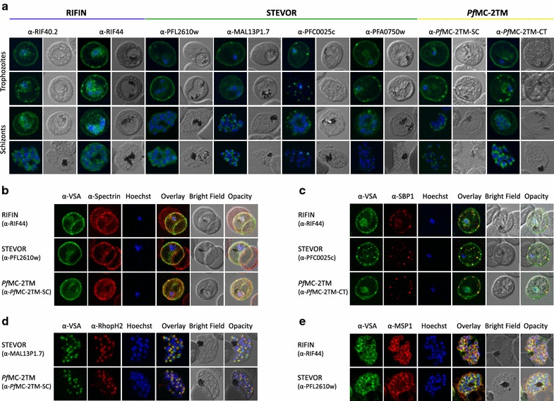
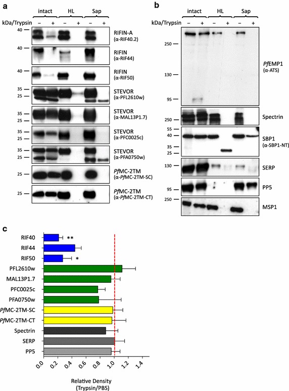
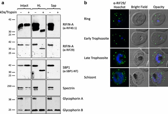
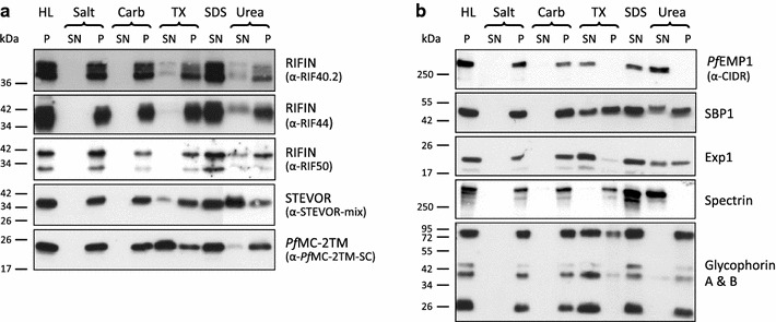
Methods: This study presents a detailed comparative characterization of the localization, membrane topology and extraction profile across the life cycle of various members of these protein families employing confocal microscopy, immunoelectron microscopy and immunoblots.

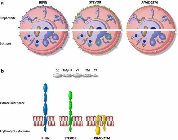
Results: The presented data reveal a clear association of variants of the RIFIN, STEVOR and PfMC-2TM proteins with the host cell membrane and topological studies indicate that the semi-conserved N-terminal region of RIFINs and some STEVOR proteins is exposed at the erythrocyte surface. At the Maurer's clefts, the semi-conserved N-terminal region as well as the variable stretch of RIFINs appears to point to the lumen away from the erythrocyte cytoplasm. These results challenge the previously proposed two transmembrane topology model for the RIFIN and STEVOR protein families and suggest that only one hydrophobic region spans the membrane. In contrast, PfMC-2TM proteins indeed seem to be anchored by two hydrophobic stretches in the host cell membrane exposing just a few, variable amino acids at the surface of the host cell.

Conclusion: Together, the host cell surface exposure and topology of RIFIN and STEVOR proteins suggests members of these protein families may indeed be involved in immune evasion of the infected erythrocyte, whereas members of the PfMC-2TM family seem to bear different functions in parasite biology.


Figures

Figure 1
Figure 1
Localization of small VSA in infected erythrocytes using confocal immunofluorescence analysis. a Asexual parasites of the 3D7 parasite clone at the trophozoite and schizont stages were fixed with methanol and small VSA localization was visualized using antibodies directed against RIFIN (α-RIF40.2, α-RIF44), STEVOR (α-PFL2610w, α-MAL13P1.7, α-PFC0025c, α-PFA0750w) and PfMC-2TM (α-PfMC-2TM-SC, α-PfMC-2TM-CT) proteins (green). Nuclei were stained with Hoechst33342 (blue). b Co-localization of α-RIF44, STEVOR α-PFL2610w and α-PfMC-2TM-SC (green) with human spectrin (red). c Co-localization of α-RIF44, STEVOR α-PFC0025c and α-PfMC-2TM-CT (green) with SBP1 (red). d Co-localization of STEVOR α-MAL13P1.7 or α-PfMC-2TM-SC (green) with the rhoptry marker RhopH2 (red). e Co-localization of α-RIF44 and STEVOR α-PFL2610w (green) with the merozoite surface protein MSP1 (red).
Figure 2
Figure 2
Immunoelectron microscopy of saponin permeabilized IE to confirm PfMC-2TM presence at the erythrocyte membrane. a, b A pre-embedding staining protocol was applied to analyse PfMC-2TM membrane association by immunoelectron microscopy. Trophozoite IE were permeabilized with saponin and incubated with the immune serum rabbit α-PfMC-2TM-CT (I) or the respective pre-immune serum (PI). Recognized proteins are visualized with 10 nm gold particles. Different sections are shown depicting PfMC-2TM association with the erythrocyte membrane (a) and with Maurer’s clefts (b). c 20 randomly selected infected erythrocytes were quantified for their gold particle localizations, which are divided into the sections total cell (total), erythrocyte membrane (EM), Maurer’s clefts (MC), parasite membrane/parasitophorous vacuole membrane (PM/PVM) and other localization (others). Data are presented as mean ± SEM. Statistical analyses were done with an unpaired t test. Significant differences between α-PfMC-2TM-CT and pre-immune serum were observed for total cells (p = 0.0003), an erythrocyte membrane association (p = 0.002) and labelling of the Maurer’s clefts (p = 0.013).
Figure 3
Figure 3
Topology of small VSA at the host cell membrane. a, b Western Blot analysis after protease treatment. MACS enriched infected erythrocytes (mostly schizonts) of the 3D7 strain were left intact (intact), subjected to hypotonic lysis (HL) or permeabilized with saponin (Sap) and subsequently also either treated with trypsin (+) or mock-treated with PBS (−). All samples were solubilized in SDS sample buffer, separated by SDS-PAGE and analysed by immunoblotting. Equivalents of 1 × 107 cells were loaded in each lane. The blots were probed with α-VSA sera as indicated (a). As controls, α-ATS antibodies against the acidic terminal segment of PfEMP1 proteins, α-Spectrin serum against the erythrocyte cytoskeleton protein spectrin and α-SBP1-NT antibodies directed against the N-terminal domain of the MC resident skeleton binding protein SBP1 as well as α-MSP1, α-SERP and α-PP5 were used (b). c Quantification of the small VSA-specific immunoblot signals after surface trypsinization of infected erythrocytes by densitometry. Three replicate experiments were quantified and data are presented as relative density from trypsin treated versus PBS treated samples adjusted to SBP1 (red line). The control proteins spectrin, SERP and PP5 are shown in grey as a reference. T test, **p < 0.01, *p < 0.05.
Figure 4
Figure 4
Membrane topology of RIFINs at the Maurer’s clefts. a Trophozoites of the rosetting strain FCR3S1.2 were analysed by protease protection assay. Intact cells (lanes 1 and 2) or cells permeabilized by hypotonic lysis (HL) or saponin (Sap) were treated with trypsin (+) or left untreated (−). Proteins of 1 × 107 cells were separated by SDS-PAGE and visualized by immunoblotting using α-RIF40.1 and α-RIF29 as well as α-SBP1-NT, α-Spectrin, and α-Glycophorin A/B antisera to control experimental performance. b The α-RIF29 antiserum stains mainly Maurer’s clefts in immunofluorescence assay of 3D7 parasites at different stages (green). Nuclei were stained with Hoechst33342 (blue).
Figure 5
Figure 5
Membrane extraction profile of small VSA proteins. Trophozoite infected erythrocytes of the 3D7 strain were lysed by hypotonic lysis (HL) and the pellet fraction was separated by centrifugation. The membrane pellet fraction was extracted with salt (Salt), carbonate (Carb), Triton X-100 (TX), SDS (SDS) or urea (Urea) containing buffers, respectively, and separated into a soluble (SN) and an insoluble (P) fraction by centrifugation. Equivalents of 1 × 107 cells were loaded in each lane. a Proteins were visualized by western blot analysis with the antibodies α-RIF40.2, α-RIF44, α-RIF50, a mixture of all α-STEVOR sera and α-PfMC-2TM-SC. b To control extraction performances, blots were probed with the antibodies α-CIDR, α-ATS, α-SBP1, α-Exp1, α-Spectrin and α-Glycophorin A/B directed against proteins with known solubilities.
Figure 6
Figure 6
Model of the localization and membrane topology of RIFIN, STEVOR and PfMC-2TM proteins. a Localization of RIFIN, STEVOR and PfMC-2TM proteins during parasite development in the erythrocyte. In trophozoite-infected erythrocytes, RIFIN (blue), STEVOR (green) and PfMC-2TM (yellow) proteins were transported to the Maurer’s clefts (MC) and most of them onwards to the erythrocyte membrane (EM). In schizonts, all small VSAs were observed at the apical tip of merozoites. Particular STEVOR variants were found at the rhoptries and others were detected at the merozoites membrane. b Proposed transmembrane topology for RIFIN, STEVOR and PfMC-2TM proteins at the EM. RIFIN and STEVOR proteins are diminished upon surface trypsinization using antisera specific for the semi-conserved N-terminal region of the proteins. Hence, a one transmembrane topology is most likely for RIFIN and a subpopulation of STEVOR proteins, which extend their semi-conserved region into the extracellular space. On the contrary, the semi-conserved as well as the C-terminal domain of PfMC-2TM proteins inserted into the erythrocyte membrane were protected from protease cleavage. Consequently, PfMC-2TM proteins seem to be inserted by two transmembrane domains and expose just a few amino acids at the surface of IE. AC apical complex, CT C-terminal domain, EM erythrocyte membrane, FV Food vacuole, HR hydrophobic region, MC Maurer’s clefts, N nucleus, PM plasma membrane, PVM parasitophorous vacuole membrane, SC semi-conserved region, TM transmembrane domain, VR variable region.

Similar articles

Cited by

References

    1. Bruce-Chwatt LJ. A longitudinal survey of natural malaria infection in a group of West African adults. West Afr Med J. 1963;12:199–217. - PubMed
    1. McGregor IA. Mechanisms of acquired immunity and epidemiological patterns of antibody responses in malaria in man. Bull World Health Organ. 1974;50:259–266. - PMC - PubMed
    1. Ferreira MU, da Silva Nunes M, Wunderlich G. Antigenic diversity and immune evasion by malaria parasites. Clin Diagn Lab Immunol. 2004;11:987–995. - PMC - PubMed
    1. Gardner MJ, Hall N, Fung E, White O, Berriman M, Hyman RW, et al. Genome sequence of the human malaria parasite Plasmodium falciparum. Nature. 2002;419:498–511. doi: 10.1038/nature01097. - DOI - PMC - PubMed
    1. Roberts DJ, Craig AG, Berendt AR, Pinches R, Nash G, Marsh K, et al. Rapid switching to multiple antigenic and adhesive phenotypes in malaria. Nature. 1992;357:689–692. doi: 10.1038/357689a0. - DOI - PMC - PubMed

Publication types

MeSH terms

Substances

LinkOut - more resources