

Baseline and longitudinal grey matter changes in newly diagnosed Parkinson's disease: ICICLE-PD study
- PMID: 26173861
- PMCID: PMC4671477
- DOI: 10.1093/brain/awv211
Baseline and longitudinal grey matter changes in newly diagnosed Parkinson's disease: ICICLE-PD study
Abstract
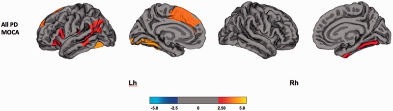
Mild cognitive impairment in Parkinson's disease is associated with progression to dementia (Parkinson's disease dementia) in a majority of patients. Determining structural imaging biomarkers associated with prodromal Parkinson's disease dementia may allow for the earlier identification of those at risk, and allow for targeted disease modifying therapies. One hundred and five non-demented subjects with newly diagnosed idiopathic Parkinson's disease and 37 healthy matched controls had serial 3 T structural magnetic resonance imaging scans with clinical and neuropsychological assessments at baseline, which were repeated after 18 months. The Movement Disorder Society Task Force criteria were used to classify the Parkinson's disease subjects into Parkinson's disease with mild cognitive impairment (n = 39) and Parkinson's disease with no cognitive impairment (n = 66). Freesurfer image processing software was used to measure cortical thickness and subcortical volumes at baseline and follow-up. We compared regional percentage change of cortical thinning and subcortical atrophy over 18 months. At baseline, cases with Parkinson's disease with mild cognitive impairment demonstrated widespread cortical thinning relative to controls and atrophy of the nucleus accumbens compared to both controls and subjects with Parkinson's disease with no cognitive impairment. Regional cortical thickness at baseline was correlated with global cognition in the combined Parkinson's disease cohort. Over 18 months, patients with Parkinson's disease with mild cognitive impairment demonstrated more severe cortical thinning in frontal and temporo-parietal cortices, including hippocampal atrophy, relative to those with Parkinson's disease and no cognitive impairment and healthy controls, whereas subjects with Parkinson's disease and no cognitive impairment showed more severe frontal cortical thinning compared to healthy controls. At baseline, Parkinson's disease with no cognitive impairment converters showed bilateral temporal cortex thinning relative to the Parkinson's disease with no cognitive impairment stable subjects. Although loss of both cortical and subcortical volume occurs in non-demented Parkinson's disease, our longitudinal analyses revealed that Parkinson's disease with mild cognitive impairment shows more extensive atrophy and greater percentage of cortical thinning compared to Parkinson's disease with no cognitive impairment. In particular, an extension of cortical thinning in the temporo-parietal regions in addition to frontal atrophy could be a biomarker in therapeutic studies of mild cognitive impairment in Parkinson's disease for progression towards dementia.
Keywords: Parkinson’s disease; dementia; neurodegeneration; neuroimaging.
© The Author (2015). Published by Oxford University Press on behalf of the Guarantors of Brain.
Figures


Similar articles
-
Tracking Cortical Changes Throughout Cognitive Decline in Parkinson's Disease.Mov Disord. 2020 Nov;35(11):1987-1998. doi: 10.1002/mds.28228. Epub 2020 Sep 4. Mov Disord. 2020. PMID: 32886420
-
Progression of subcortical atrophy in mild Parkinson's disease and its impact on cognition.Eur J Neurol. 2017 Feb;24(2):341-348. doi: 10.1111/ene.13205. Epub 2016 Dec 10. Eur J Neurol. 2017. PMID: 27943468
-
Mild cognitive impairment is linked with faster rate of cortical thinning in patients with Parkinson's disease longitudinally.Brain. 2014 Apr;137(Pt 4):1120-9. doi: 10.1093/brain/awu036. Epub 2014 Mar 10. Brain. 2014. PMID: 24613932
-
Progression of grey and white matter brain damage in Parkinson's disease: a critical review of structural MRI literature.J Neurol. 2021 Sep;268(9):3144-3179. doi: 10.1007/s00415-020-09863-8. Epub 2020 May 6. J Neurol. 2021. PMID: 32378035 Review.
-
Evolution of cognitive dysfunction in an incident Parkinson's disease cohort.Brain. 2007 Jul;130(Pt 7):1787-98. doi: 10.1093/brain/awm111. Epub 2007 May 29. Brain. 2007. PMID: 17535834 Review.
Cited by
-
Meynert nucleus-related cortical thinning in Parkinson's disease with mild cognitive impairment.Quant Imaging Med Surg. 2021 Apr;11(4):1554-1566. doi: 10.21037/qims-20-444. Quant Imaging Med Surg. 2021. PMID: 33816191 Free PMC article.
-
Cortical thickness as predictor of response to exercise in people with Parkinson's disease.Hum Brain Mapp. 2021 Jan;42(1):139-153. doi: 10.1002/hbm.25211. Epub 2020 Oct 9. Hum Brain Mapp. 2021. PMID: 33035370 Free PMC article. Clinical Trial.
-
Interaction Between Neuropsychiatric Symptoms and Cognitive Performance in Parkinson's Disease: What Do Clinical and Neuroimaging Studies Tell Us?Curr Neurol Neurosci Rep. 2018 Oct 15;18(12):91. doi: 10.1007/s11910-018-0907-6. Curr Neurol Neurosci Rep. 2018. PMID: 30324260 Review.
-
A divergent breakdown of neurocognitive networks in Parkinson's Disease mild cognitive impairment.Hum Brain Mapp. 2019 Aug 1;40(11):3233-3242. doi: 10.1002/hbm.24593. Epub 2019 Apr 1. Hum Brain Mapp. 2019. PMID: 30938027 Free PMC article.
-
Progressive brain atrophy in Parkinson's disease patients who convert to mild cognitive impairment.CNS Neurosci Ther. 2020 Jan;26(1):117-125. doi: 10.1111/cns.13188. Epub 2019 Jul 6. CNS Neurosci Ther. 2020. PMID: 31278861 Free PMC article.
References
-
- Aarsland D, Andersen K, Larsen JP, Lolk A. Prevalence and characteristics of dementia in parkinson disease. Arch Neurol 2003; 60: 387–92. - PubMed
-
- Aarsland D, Larsen JP, Karlsen K, Lim NG, Tandberg E. Mental symptoms in Parkinson’s disease are important contributors to caregiver distress. Int J Geriatr Psychiatry 1999; 14: 866–74. - PubMed
-
- Burton EJ, McKeith IG, Burn DJ, O’Brien JT. Brain atrophy rates in Parkinson’s disease with and without dementia using serial magnetic resonance imaging. Mov Disord 2005; 20: 1571–6. - PubMed
Publication types
MeSH terms
Grants and funding
LinkOut - more resources
Full Text Sources
Other Literature Sources
Medical
