. 2015 Jul;115 Suppl 1(Suppl 1):i66-i76.
doi: 10.1093/bja/aev114.

Age-dependency of sevoflurane-induced electroencephalogram dynamics in children

Affiliations

Age-dependency of sevoflurane-induced electroencephalogram dynamics in children

O Akeju et al. Br J Anaesth. 2015 Jul.

Abstract

Background: General anaesthesia induces highly structured oscillations in the electroencephalogram (EEG) in adults, but the anaesthesia-induced EEG in paediatric patients is less understood. Neural circuits undergo structural and functional transformations during development that might be reflected in anaesthesia-induced EEG oscillations. We therefore investigated age-related changes in the EEG during sevoflurane general anaesthesia in paediatric patients.

Methods: We analysed the EEG recorded during routine care of patients between 0 and 28 yr of age (n=54), using power spectral and coherence methods. The power spectrum quantifies the energy in the EEG at each frequency, while the coherence measures the frequency-dependent correlation or synchronization between EEG signals at different scalp locations. We characterized the EEG as a function of age and within 5 age groups: <1 yr old (n=4), 1-6 yr old (n=12), >6-14 yr old (n=14), >14-21 yr old (n=11), >21-28 yr old (n=13).

Results: EEG power significantly increased from infancy through ∼6 yr, subsequently declining to a plateau at approximately 21 yr. Alpha (8-13 Hz) coherence, a prominent EEG feature associated with sevoflurane-induced unconsciousness in adults, is absent in patients <1 yr.

Conclusions: Sevoflurane-induced EEG dynamics in children vary significantly as a function of age. These age-related dynamics likely reflect ongoing development within brain circuits that are modulated by sevoflurane. These readily observed paediatric-specific EEG signatures could be used to improve brain state monitoring in children receiving general anaesthesia.

Keywords: electroencephalography; pediatric; sevoflurane.


Figures

Fig 1
Fig 1
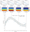
Trends in spectra, spectrograms, and total power with age from 0 to 28 yr old. (ad) Representative frontal EEG spectra illustrating that slow (0.1–1 Hz), and delta (1–4 Hz) oscillations are present in all patients during general anaesthesia, maintained solely with sevoflurane. Alpha (8–12 Hz) oscillations appear to emerge after 1 yr old. (eh) Representative frontal EEG spectrograms illustrating that slow (0.1–1 Hz), and delta (1–4 Hz) oscillations are present in all patients during general anaesthesia. Alpha (8–12 Hz) oscillations appear to emerge after 1 yr of age. (i) Total EEG power (1–50 Hz) for each subject, plotted as a function of age. The total EEG power exhibited an increase from infancy, peaked at approximately 5–8 yr old, and subsequently declined with increasing age. The green line represents a fourth degree polynomial regression model describing the relationship between age and EEG power. The shaded bounds represent the 95% confidence bounds of this regression model.
Fig 2
Fig 2
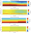
Age varying spectrograms and coherograms during sevoflurane general anaesthesia. (a) An age varying (1–28 yr) spectrogram representation of the EEG, showing that even though the EEG structure appears qualitatively preserved for all age ranges (presence of slow, delta, theta, and alpha oscillations), the power of these oscillations change as a function of age. (b) An age varying (1–28 yr) coherogram illustrating stably maintained, prominent alpha oscillation coherence. (c) An age varying spectrogram based representation of the EEG showing the relative absence of a well-defined alpha oscillation band at less than 1 yr of age. At approximately 1 yr of age the alpha oscillations become prominent. Slow, delta and theta oscillations are present from 0–1.5 yr. (d) An age varying coherogram illustrating the absence of alpha oscillation coherence, and slow delta band coherence, at less than 1 yr of age. This EEG dynamic appears at >1 yr of age, and is inversely correlated to slow oscillation coherence.
Fig 3
Fig 3
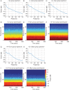
Median spectra and spectrograms of age groups. (a, d) Group 1 (<1 yr old). Both the power spectra and the group spectrogram show large power in the slow, delta and theta frequency bands. A prominent alpha oscillation structure is noticeably absent. (b, e) Group 2 (1–6 yr old). Both the power spectra and the group spectrogram show large power in the slow, delta, theta and alpha frequency bands. (c, f) Group 3 (>6–14 yr old). Both the power spectra and the group spectrogram show large power in the slow, delta, theta and alpha frequency bands. (g, i) Group 4 (>14–21 yr old). Both the power spectra and the group spectrogram show large power in the slow, delta, theta and alpha frequency bands. Compared with groups 2 and 3, the power in these bands is not as prominent. Power within the beta/gamma frequency band also shifts to lower levels. (h, j) Group 5 (>21–28 yr old). Both the power spectra and the group spectrogram show large power in the slow, delta, theta and alpha frequency bands. Compared with groups 2 and 3, the power in these bands is not as prominent. Power within the beta/gamma frequency band also shifts to lower levels.
Fig 4
Fig 4
Median coherence and coherograms of age groups. (a, d) Group 1 (<1 yr old). The median coherence and coherogram shows a lack of coherence in the alpha frequency band and significant coherence in the slow, and delta bands. (b, e) Group 2 (1–6 yr old). The median coherence and coherogram shows a strong coherence in the alpha frequency band. (c, f) Group 3 (>6–14 yr old). The median coherence and coherogram shows a strong coherence in the alpha frequency band. (g, i) Group 4 (>14–21 yr old). The median coherence and coherogram shows a strong coherence in the alpha frequency band. (h, j) Group 5 (>21–28 yr old). The median coherence and coherogram shows a strong coherence in the alpha frequency band.

Similar articles

Cited by

References

    1. Jevtovic-Todorovic V, Absalom AR, Blomgren K, et al. Anaesthetic neurotoxicity and neuroplasticity: an expert group report and statement based on the BJA Salzburg Seminar. Br J Anaesth 2013; 111: 143–51 - PMC - PubMed
    1. Lin EP, Soriano SG, Loepke AW. Anesthetic neurotoxicity. Anesthesiol Clin 2014; 32: 133–55 - PubMed
    1. McCann ME, Soriano SG. General anesthetics in pediatric anesthesia: influences on the developing brain. Curr Drug Targets 2012; 13: 944–51 - PMC - PubMed
    1. Wilder RT, Flick RP, Sprung J, et al. Early exposure to anesthesia and learning disabilities in a population-based birth cohort. Anesthesiology 2009; 110: 796–804 - PMC - PubMed
    1. Sanders RD, Hassell J, Davidson AJ, Robertson NJ, Ma D. Impact of anaesthetics and surgery on neurodevelopment: an update. Br J Anaesth 2013; 110(Suppl 1): i53–72 - PMC - PubMed

Publication types