

Ageing induced vascular smooth muscle cell senescence in atherosclerosis
- PMID: 26174609
- PMCID: PMC4933105
- DOI: 10.1113/JP270923
Ageing induced vascular smooth muscle cell senescence in atherosclerosis
Abstract
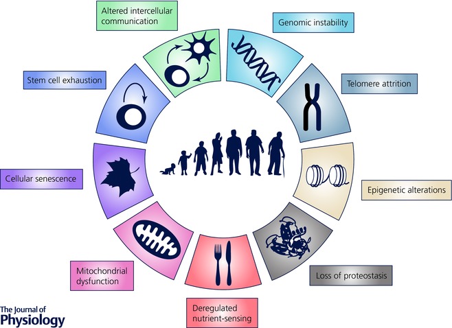
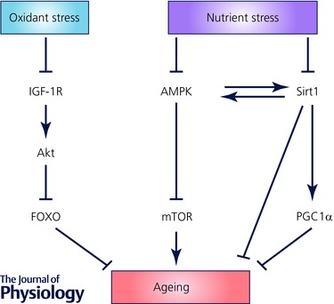
Atherosclerosis is a disease of ageing in that its incidence and prevalence increase with age. However, atherosclerosis is also associated with biological ageing, manifest by a number of typical hallmarks of ageing in the atherosclerotic plaque. Thus, accelerated biological ageing may be superimposed on the effects of chronological ageing in atherosclerosis. Tissue ageing is seen in all cells that comprise the plaque, but particularly in vascular smooth muscle cells (VSMCs). Hallmarks of ageing include evidence of cell senescence, DNA damage (including telomere attrition), mitochondrial dysfunction, a pro-inflammatory secretory phenotype, defects in proteostasis, epigenetic changes, deregulated nutrient sensing, and exhaustion of progenitor cells. In this model, initial damage to DNA (genomic, telomeric, mitochondrial and epigenetic changes) results in a number of cellular responses (cellular senescence, deregulated nutrient sensing and defects in proteostasis). Ultimately, ongoing damage and attempts at repair by continued proliferation overwhelm reparative capacity, causing loss of specialised cell functions, cell death and inflammation. This review summarises the evidence for accelerated biological ageing in atherosclerosis, the functional consequences of cell ageing on cells comprising the plaque, and the causal role that VSMC senescence plays in atherogenesis.
© 2015 The Authors. The Journal of Physiology © 2015 The Physiological Society.
Figures
References
-
- Aavik E, Lumivuori H, Leppanen O, Wirth T, Hakkinen SK, Brasen JH, Beschorner U, Zeller T, Braspenning M, van Criekinge W, Makinen K & Yla‐Herttuala S (2015). Global DNA methylation analysis of human atherosclerotic plaques reveals extensive genomic hypomethylation and reactivation at imprinted locus 14q32 involving induction of a miRNA cluster. Eur Heart J 36, 993–1000. - PubMed
-
- Acosta JC, Banito A, Wuestefeld T, Georgilis A, Janich P, Morton JP, Athineos D, Kang TW, Lasitschka F, Andrulis M, Pascual G, Morris KJ, Khan S, Jin H, Dharmalingam G, Snijders AP, Carroll T, Capper D, Pritchard C, Inman GJ, Longerich T, Sansom OJ, Benitah SA, Zender L & Gil J (2013). A complex secretory program orchestrated by the inflammasome controls paracrine senescence. Nat Cell Biol 15, 978–990. - PMC - PubMed
-
- Acosta JC, O'Loghlen A, Banito A, Guijarro MV, Augert A, Raguz S, Fumagalli M, Da Costa M, Brown C, Popov N, Takatsu Y, Melamed J, d'Adda di Fagagna F, Bernard D, Hernando E & Gil J (2008). Chemokine signaling via the CXCR2 receptor reinforces senescence. Cell 133, 1006–1018. - PubMed
-
- Allard D, Figg N, Bennett MR & Littlewood TD (2008). Akt regulates the survival of vascular smooth muscle cells via Inhibition of FoxO3a and GSK3. J Biol Chem 283, 19739–19747. - PubMed
-
- Aubert G & Lansdorp PM (2008). Telomeres and aging. Physiol Rev 88, 557–579. - PubMed
Publication types
MeSH terms
Grants and funding
LinkOut - more resources
Full Text Sources
Other Literature Sources
Medical