Review
. 2015 Sep;8(5):959-68.
doi: 10.1038/mi.2015.63. Epub 2015 Jul 15.

Wound repair: role of immune-epithelial interactions

Affiliations
Review

Wound repair: role of immune-epithelial interactions

G Leoni et al. Mucosal Immunol. 2015 Sep.

Abstract

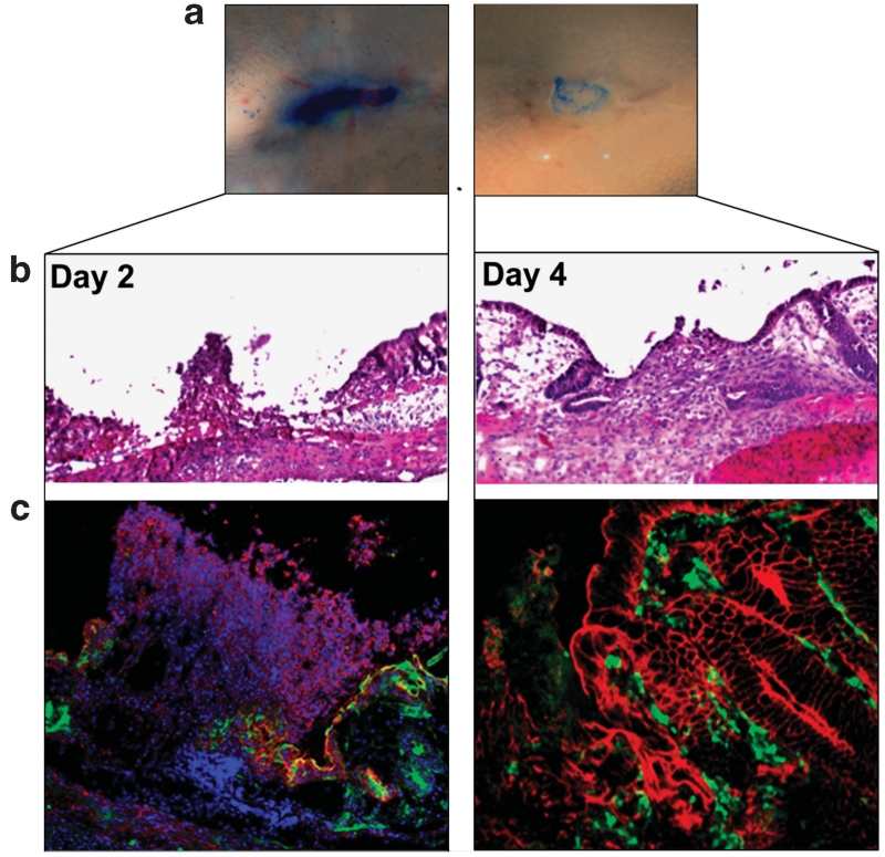
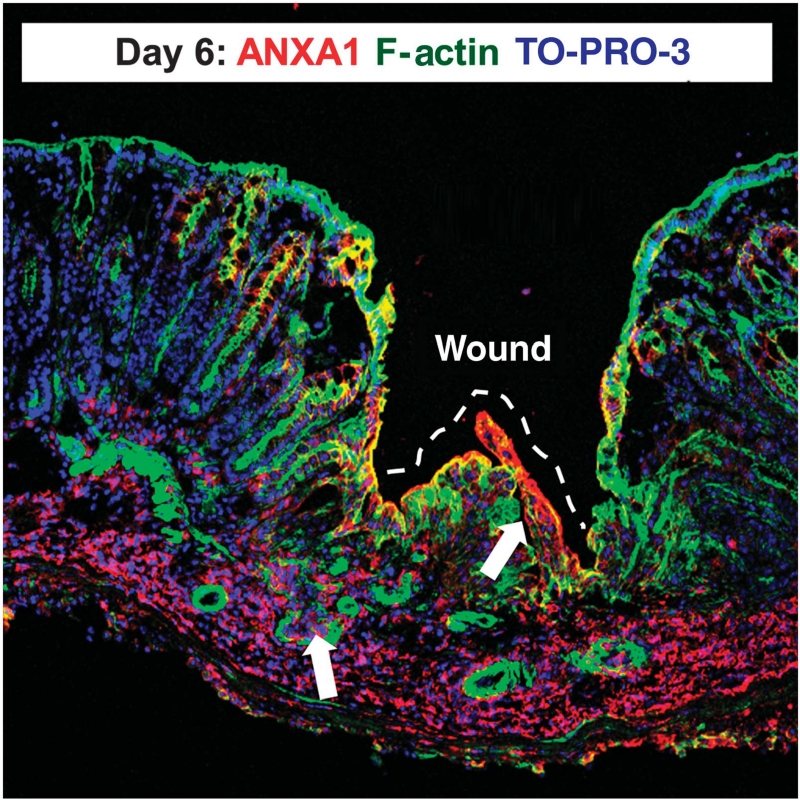
The epithelium serves as a highly selective barrier at mucosal surfaces. Upon injury, epithelial wound closure is orchestrated by a series of events that emanate from the epithelium itself as well as by the temporal recruitment of immune cells into the wound bed. Epithelial cells adjoining the wound flatten out, migrate, and proliferate to rapidly cover denuded surfaces and re-establish mucosal homeostasis. This process is highly regulated by proteins and lipids, proresolving mediators such as Annexin A1 protein and resolvins released into the epithelial milieu by the epithelium itself and infiltrating innate immune cells including neutrophils and macrophages. Failure to achieve these finely tuned processes is observed in chronic inflammatory diseases that are associated with non-healing wounds. An improved understanding of mechanisms that mediate repair is important in the development of therapeutics aimed to promote mucosal wound repair.


Figures

Figure 1
Figure 1
Spatio-temporal control of wound closure. The initial phase of wound closure is characterized by an influx of neutrophils (day 1) followed by increased presence of macrophages (days 2–4) at the injured site. The inflammatory response is associated with re-epithelialization of the mucosa that is achieved by migration and subsequent proliferation of epithelial cells.
Figure 2
Figure 2
Resealing intestinal mucosal wounds. (a) Photographs obtained by colonoscopy of mice with resealing biopsy-induced colonic wounds on days 2 and 4 after injury highlighted with methylene blue. (b) Histology of intestinal mucosal wounds stained with hematoxylin and eosin. (c) Representative immunofluorescence confocal images showing neutrophils (visualized by anti-Ly6G staining, pink) and macrophages (visualized by anti-F4/80 staining, green) in intestinal mucosal wounds on days 2 and 4 post wounding.
Figure 3
Figure 3
Mucosal ANXA1 expression is increased in repairing intestinal mucosal wounds. Representative image of murine colonic mucosa 2 days after biopsy-induced injury. Frozen sections were stained with antibodies against ANXA1 (red), F-actin (phalloidin, green), and nuclei (TO-PRO-3, blue). Arrows show ANXA1 expression by epithelial and immune cells in the injured mucosa.
Figure 4
Figure 4
Acute and chronic wounds. During acute wound closure a plethora of signaling molecules emanating from the epithelium and immune cells coordinate wound repair (upper panel). In addition to cytokines, chemokines and growth factors, a distinct set of proresolving mediators such as protectins, resolvins, maresins, and proteins such as ANXA1 orchestrate wound repair. Chronic wounds (lower panel) are characterized by the presence of a high number of inflammatory cells (neutrophils) that generate a predominantly proinflammatory milieu, characterized by high concentrations of reactive oxygen species (ROS) as well as matrix metalloproteinases (MMPs) that in turn lead to destruction of the extracellular matrix, disruption of epithelial contacts, and result in further invasion of pathogens. HIF-1, hypoxia-inducible factor-1; MPO, myeloperoxidase.

References

    1. Peterson LW, Artis D. Intestinal epithelial cells: regulators of barrier function and immune homeostasis. Nat. Rev. Immunol. 2014;14:141–153. - PubMed
    1. Gurtner GC, Werner S, Barrandon Y, Longaker MT. Wound repair and regeneration. Nature. 2008;453:314–321. - PubMed
    1. Wong JW, et al. Wound healing in oral mucosa results in reduced scar formation as compared with skin: evidence from the red Duroc pig model and humans. Wound Repair Regen. 2009;17:717–729. - PubMed
    1. Nusrat A, Delp C, Madara JL. Intestinal epithelial restitution. Characterization of a cell culture model and mapping of cytoskeletal elements in migrating cells. J. Clin. Invest. 1992;89:1501–1511. - PMC - PubMed
    1. Yang HW, et al. Cooperative activation of PI3K by Ras and Rho family small GTPases. Mol. Cell. 2012;47:281–290. - PMC - PubMed