. 2015 Nov;62(5):1466-79.
doi: 10.1002/hep.27987. Epub 2015 Aug 28.

NUMB phosphorylation destabilizes p53 and promotes self-renewal of tumor-initiating cells by a NANOG-dependent mechanism in liver cancer

Affiliations

NUMB phosphorylation destabilizes p53 and promotes self-renewal of tumor-initiating cells by a NANOG-dependent mechanism in liver cancer

Hifzur R Siddique et al. Hepatology. 2015 Nov.

Abstract

Stem cell populations are maintained through self-renewing divisions in which one daughter cell commits to a particular fate whereas the other retains the multipotent characteristics of its parent. The NUMB, a tumor suppressor, in conjunction with another tumor-suppressor protein, p53, preserves this property and acts as a barrier against deregulated expansion of tumor-associated stem cells. In this context, NUMB-p53 interaction plays a crucial role to maintain the proper homeostasis of both stem cells, as well as differentiated cells. Because the molecular mechanism governing the assembly and stability of the NUMB-p53 interaction/complex are poorly understood, we tried to identify the molecule(s) that govern this process. Using cancer cell lines, tumor-initiating cells (TICs) of liver, the mouse model, and clinical samples, we identified that phosphorylations of NUMB destabilize p53 and promote self-renewal of TICs in a pluripotency-associated transcription factor NANOG-dependent manner. NANOG phosphorylates NUMB by atypical protein kinase C zeta (aPKCζ), through the direct induction of Aurora A kinase (AURKA) and the repression of an aPKCζ inhibitor, lethal (2) giant larvae. By radioactivity-based kinase activity assays, we showed that NANOG enhances kinase activities of both AURKA and aPKCζ, an important upstream process for NUMB phosphorylation. Phosphorylation of NUMB by aPKCζ destabilizes the NUMB-p53 interaction and p53 proteolysis and deregulates self-renewal in TICs.

Conclusion: Post-translational modification of NUMB by the NANOG-AURKA-aPKCζ pathway is an important event in TIC self-renewal and tumorigenesis. Hence, the NANOG-NUMB-p53 signaling axis is an important regulatory pathway for TIC events in TIC self-renewal and liver tumorigenesis, suggesting a therapeutic strategy by targeting NUMB phosphorylation. Further in-depth in vivo and clinical studies are warranted to verify this suggestion.


Conflict of interest statement

Disclosures of Potential Conflict of Interest: Authors have nothing to disclose.

Figures

Figure 1
Figure 1. NUMB phosphorylations and p53 levels are linked to NANOG level and in the Tumor Initiating Cells (TICs) and Clinical Tissues
(A) Immunoblot analysis of lysates prepared from patient HCC tissue specimens and matched, non-cancerous tissues using the indicated antisera. (Bi) Representative photomicrographs showing the expression of NANOG and pNUMB (Ser-265) in human HCCs and adjacent non-cancerous tissues as assessed by an immunofluorescence assay. The merged lane shows the co-localization of 2 proteins. DAPI was used as a nuclear staining control. (Bii) Comparison of Immuno Reactivity Score (IRS, the product of a percent of staining and intensity of staining) in tumor vs. non-tumor tissues. (C) Immunoblot analysis of lysates prepared from human hepatocytes stably expressed or constitutively active TLR4 (caTLR4). The lysates were analyzed using the indicated antisera. (D) Immunoblot analysis of lysates prepared from patient HCC tissue specimens and matched, non-cancerous tissues using the indicated antisera. (Ei) Photomicrographs represent immunostaining of NANOG and p53 in liver specimens of liver cancer patients. formula image 
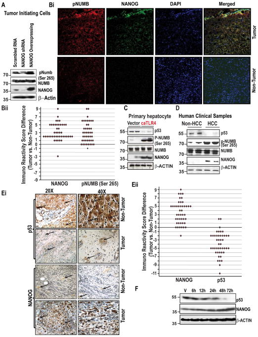
Arrows indicate staining for respective protein. Magnifications X20 and X40. (Eii) Comparison of Immuno Recativity Score (IRS, product of percent of staining and intensity of staining) in tumor vs. non-tumor tissues. (F) Figure represents the level of NANOG and p53 proteins in NANOG transfected Huh7 cells as assessed by immunoblot analysis.
Figure 2
Figure 2. NANOG modulates LGL2 and aPKCζ, a NUMB kinase, expression and activity
(A) NANOG overexpression promotes phosphorylation of aPKCζ. (B) Read density for NANOG ChIP-seq libraries at the Lgl-2 locus in CD133+ TICs and CD133 controls. A red bar representing a significantly enriched NANOG binding site detected at the Lgl-2 locus is shown. Representations of the annotated mRNA and coding sequencing (CDS) for Lgl-2 are shown at the top. (Ci) NANOG represses LGL-2 expression as measured by qRT-PCR. Error bars represent the SD from at least three independent biological replicates (*P < 0.05). (Cii) NANOG inhibits expression levels of LGL-2 protein. (D) Silencing of NANOG transcriptionally activates LGL-2 promoter. Huh7 cells stably overexpressing NANOG or scrambled shRNA, or sh-NANOG were transduced with the LGL-2 promoter sequences placed upstream of a firefly luciferase reporter gene. Promoter activity is displayed as relative light units (RLU) normalized to the activity of cotransfected Renilla luciferase. Error bars represent the SD from at least three independent biological replicates (**P < 0.05). (Ei–ii) The histogram represents the effect of NANOG-silenced and -overexpression of the kinase activity of aPKCζ in Huh7 cells (Ei) and TICs (Eii). (Fi–ii) The histogram represents the effect of Lipopolysaccharides on aPKCζ kinase activity Huh7 cells (Fi) and TICs (Fii). Each bar in the histogram represents mean ± SD of three independent experiments, * represents P < 0.05.
Figure 3
Figure 3. NANOG acts as a transcriptional activator of AURKA and modulates kinase activity
(A) ChIP-qPCR demonstrates direct association of NANOG at the AURKA promoter. Huh7, HepG2 and Hep3B cells stably expressing NANOG or harboring a non-targeting, scrambled shRNA, or NANOG-targeting shRNA were subjected to Chromatin immunoprecipitation using ChIP grade NANOG antisera or isotype-matched control IgG. Immunoprecipitated chromatin was analyzed by qPCR using primer sets designed to amplify the indicated regions. (B) AURKA promoter reporter assay. Huh7 cells were transfected with the indicated promoter-reporter constructs. (C) Mutation of NANOG-binding sites reduces levels of transactivation. Promoter activity is expressed as relative light units (RLU) normalized to the activity of cotransfected Renilla luciferase. Error bars represent the SD from at least three independent biological replicates (*P < 0.05). (Di–ii) Figures represent the effect of NANOG-silenced and overexpression of the kinase activity of AURKA in Huh7 cells (Di) and TICs (Dii). (Ei–ii) The histogram represents the effect of Lipopolysaccharides (LPS) on AURKA activity Huh7 cells (Ei) and TICs (Eli). Each bar in the histogram represents mean ± SD of three independent experiments, * represents P < 0.05.
Figure 4
Figure 4. Validation of AURKA and LGL-2 expression in clinical samples
(A–B) Representative photomicrographs showing the expression of NANOG, AURKA, LGL2 and pNUMB (Ser-265) in human HCCs and adjacent non-cancerous tissues as assessed by an immunofluorescence assay. The merged lane shows the co-localization of 2 proteins. DAPI was used as a nuclear staining control. (C) Comparison of Immuno Reactivity Score (IRS, the product of a percent of staining and intensity of staining) in tumor vs. non-tumor tissues p<0.05, n=45. (D) CD133+ and CD133− cells were transfected with empty vector or AURKA expression vectors and were resolved by SDS-PAGE followed by immunoblotting using the indicated antibodies.
Figure 5
Figure 5. NANOG triggers destabilization of the NUMB-p53 complex through aPKCζ-mediated phosphorylation of NUMB. NANOG induces the proteolysis of p53 in TICs, and its expression is inversely related to p53 in cells and clinical samples
(Ai) CD133+ TICs were transfected with empty vector or the indicated combinations of MYC-p53 and NANOG, followed by exposure to Nutlin-3 for 24h. Cell lysates were immunoprecipitated using anti-NUMB antibody and analyzed by SDS-PAGE and immunoblotting. (Aii) TICs were transfected with MYC-p53 and either empty vector or increasing amounts of CA-aPKCζ. Following exposure to Nutlin-3, lysates were prepared and subjected to immunoprecipitation using an anti-NUMB antibody. (B) Murine TICs were transfected with MYC-p53 together with empty vector, dominant-negative aPKCζ (dn-aPKCζ), constitutively active, aPKCζ (CA-aPKCζ). Lysates were resolved by SDS-PAGE and analyzed by immunoblotting. (Ci) (Upper panel) Structures of wild type, unphosphorylatable mutant NUMB-3A and phosphomimetic mutant of NUMB-3D. PTB: phosphotyrosine-binding domain, PRR: proline-rich region. (Ci, lower panel) Interaction of MYC-p53 with Flag-NUMB variants. TICs were transfected with empty vector or NANOG, then analyzed by SDS-PAGE and immunoblotting. Flag-NUMB or the indicated phosphorylation site variants of Flag-NUMB were expressed alone or with MYC-p53, followed by lysis and immunoprecipitation using an anti-MYC antibody. Proteins were resolved by SDS-PAGE and analyzed by immunoblotting. Lysates represent 10% of the input volume used in the immunoprecipitation. (Cii) TICs were transfected with empty vector or NANOG along with the indicated variants of Flag-NUMB, followed by lysis and immunoblotting. (Di–ii) CD133+/CD49f+ murine liver TICs were stably transduced with pG13-luc (Di) or p21-luc (Dii), in the absence or presence of p53 and with increasing amounts of NANOG expression vector. Promoter activity is expressed as relative light units (RLU) normalized to the activity of cotransfected Renilla luciferase. Error bars represent the SD from at least three independent biological replicates (**P< 0.05). (Ei) Lysates prepared from CD133+/CD49f+ TICs transfected with p53, NANOG, or both expression vectors were resolved by SDS-PAGE followed by immunoblotting using the indicated antibodies. (Eii) In vitro proteolysis assay. Purified, recombinant GST-p53 was incubated for 30 minutes in buffer alone (lane 1) or in equal amounts of lysates prepared from TICs transfected with either empty vector or NANOG at the indicated ratios. (F) TICs were transfected with NANOG or empty vector, followed by exposure to Nutlin-3 (10μM) or vehicle for 24h. Cell lysates were analyzed by immunoblotting using an anti-p53 antibody. Ponceau S stain (bottom) serves as a loading control.
Figure 6
Figure 6. aPKCζ-dependent phosphorylation of the NUMB-p53 complex promotes self-renewal and tumorigenesis
(A) Conceptual model of NANOG-mediated oncogenesis in TICs. (Bi–ii) Methylcellulose colony formation assay. TICs stably expressing the indicated combinations of NANOG and Flag-NUMB were seeded in low adhesion methylcellulose media and cultured for one week, then harvested and reseeded for a total of four platings. The number of colonies that formed for each cell type is indicated. (C) Tumor formation titration assay. TICs (102–105) stably expressing the indicated transgenes or lentivirus shRNAs were implanted subcutaneously into the dorsal hind flanks of NOG mice and tumor growth was monitored for 60 days. Tumors greater than 25 mm3 and which exhibited growth progression during the course of the study were scored as positive. (Di) Tumor growth kinetics. TICs were implanted subcutaneously into NOG mice, and tumor volumes were measured on the indicated days. (Dii) Representative photomicrographs showing the expression of CD133 in cancer cells derived xenograft and TICs derived xenograft tissues as assessed by an immunostaining. formula image 
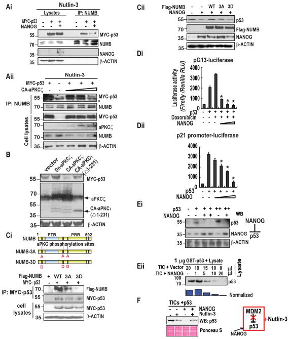
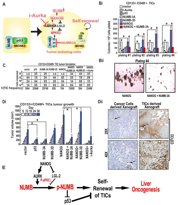
Arrows indicate staining for respective protein. Magnifications X20 and X40. (E) Postulated mechanism of self-renewal ability through NANOG-AURKA-pNUMB pathways.

References

    1. Hunter T. Signaling-2000 and beyond. Cell. 2000;100:113–27. - PubMed
    1. Schlessinger J. Cell signaling by receptor tyrosine kinases. Cell. 2000;103:211–25. - PubMed
    1. Cuesta N, Martín-Cófreces NB, Murga C, van Santen HM. Receptors, signaling networks, and disease. Sci Signal. 2011;4:mr3. - PubMed
    1. Cohen P. The twentieth century struggle to decipher insulin signalling. Nat Rev Mol Cell Biol. 2006;7:867–73. - PubMed
    1. Boulikas T. The phosphorylation connection to cancer. Int J Oncol. 1995;6:271–8. - PubMed

Publication types

MeSH terms