. 2015 Sep 11;290(37):22612-21.
doi: 10.1074/jbc.M115.661363. Epub 2015 Jul 13.

Histone core phosphorylation regulates DNA accessibility

Affiliations

Histone core phosphorylation regulates DNA accessibility

Matthew Brehove et al. J Biol Chem. .

Abstract

Nucleosome unwrapping dynamics provide transient access to the complexes involved in DNA transcription, repair, and replication, whereas regulation of nucleosome unwrapping modulates occupancy of these complexes. Histone H3 is phosphorylated at tyrosine 41 (H3Y41ph) and threonine 45 (H3T45ph). H3Y41ph is implicated in regulating transcription, whereas H3T45ph is involved in DNA replication and apoptosis. These modifications are located in the DNA-histone interface near where the DNA exits the nucleosome, and are thus poised to disrupt DNA-histone interactions. However, the impact of histone phosphorylation on nucleosome unwrapping and accessibility is unknown. We find that the phosphorylation mimics H3Y41E and H3T45E, and the chemically correct modification, H3Y41ph, significantly increase nucleosome unwrapping. This enhances DNA accessibility to protein binding by 3-fold. H3K56 acetylation (H3K56ac) is also located in the same DNA-histone interface and increases DNA unwrapping. H3K56ac is implicated in transcription regulation, suggesting that H3Y41ph and H3K56ac could function together. We find that the combination of H3Y41ph with H3K56ac increases DNA accessibility by over an order of magnitude. These results suggest that phosphorylation within the nucleosome DNA entry-exit region increases access to DNA binding complexes and that the combination of phosphorylation with acetylation has the potential to significantly influence DNA accessibility to transcription regulatory complexes.

Keywords: DNA accessibility; DNA-protein interaction; fluorescence resonance energy transfer (FRET); histone modification; histone post-translational modifications; nucleosome; small-angle X-ray scattering (SAXS).


Figures

FIGURE 1.
FIGURE 1.
Design and analysis of nucleosomes used in this study. A, nucleosome crystal structure (Protein Data Bank (PDB): 1AOI) showing the modified amino acids in blue: H3Y41, H3T45, and H3K56. For FRET measurements, Cy3 is attached at the 5′ end of the DNA molecule (green), and Cy5 is attached at H2AK119C (orange). The LexA binding site is located from the 8th to the 27th bp of the nucleosome (dark red). The region of the nucleosome containing the three PTMs is enlarged to indicate the residues' orientation relative to the DNA. B, schematic diagrams of the DNA molecules used in these studies. The green star indicates the location of the Cy3 fluorophore. C, EMSA of the nucleosomes (Nuc) used in FRET measurements.
FIGURE 2.
FIGURE 2.
Analysis of purified fully synthetic proteins H3Y41ph and H3Y41ph/K56ac. A, analytical RP-HPLC of H3Y41ph with a gradient of 32–63% acetonitrile, 0.1% TFA. Abs, absorbance. B, MALDI-TOF MS of H3Y41ph: ([MH+] m/z expected 15,280, observed 15,283; [MH2+] m/z expected 7640, observed 7640). C, analytical RP-HPLC of H3Y41ph/K56ac with a gradient of 32–63% acetonitrile, 0.1% TFA. D, MALDI-TOF MS of H3Y41ph/K56ac: ([MH+] m/z expected 15,322, observed 15,322; [MH2+] m/z expected 7661, observed 7660).
FIGURE 3.
FIGURE 3.
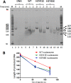
MNase digestion of WT, H3Y41E, and H3T45E nucleosomes. A, nucleosomes reconstituted with 601-207 DNA and histone octamer containing WT H3, H3Y41E or H3T45E were digested with MNase. The digestions were quenched at 0, 60, and 120 s. The length of protected DNA was analyzed on a 6% polyacrylamide gel and visualized by SYBR Gold staining. B, the length of protected DNA as a function of digestion time. The migration distance of each band (A) was measured with the one-dimensional gel analysis function of ImageQuant TL software. When compared with WT nucleosome, mutant nucleosomes are less resistant to MNase. Error bars indicate mean ± S.D.
FIGURE 4.
FIGURE 4.
WT, H3Y41E, and H3T45E nucleosomes have molecular masses consistent with a full complement of histones. To determine the molecular mass and quality of nucleosomes prior to SAXS experiments, SEC-MALS was performed. The molecular mass for each sample was calculated by the ASTRA software (Wyatt Technologies). The calculated mass for each nucleosome is within error of their theoretical mass (200 KDa), and samples are monodisperse. Because the light-scattering signal from the peak that eluted at 7–8 ml in each nucleosome sample is also present in the buffer control (data not shown), it is not due to free DNA or other contaminants. a. u., arbitrary units.
FIGURE 5.
FIGURE 5.
Substitution of H3Y41 or H3T45 with glutamic acid result in extended nucleosomes. A, experimental Rg values (in Å) for nucleosomes shown in B, at 0 mm KCl (black, left-hand side) and 50 mm KCl (blue, right-hand side). The values shown are Rg (Å) and two standard deviations, giving approximately a 95% confidence interval. B, the molecular envelope of nucleosomes containing 601-147 and WT H3, H3Y41E, or H3T45E, calculated ab initio from SAXS data taken at 0 mm KCl. The shell was superimposed onto the crystal structure of the nucleosome (PDB: 1AOI) without histone tails.
FIGURE 6.
FIGURE 6.
Fluorescence resonance energy transfer measurements of LexA binding within modified nucleosomes. A, kinetic model of a transcription factor (blue oval) binding to a partially unwrapped nucleosome and trapping the nucleosome in this partially unwrapped state. In this state, the Cy3 (green star) and Cy5 (red stars) are separated, causing low FRET. B, example emission spectra with Cy3 excitation for increasing [LexA]. Cy3 emission increases and Cy5 emission decreases as [LexA] increases, which is due to LexA binding and trapping the nucleosome in a partially unwrapped state with lower FRET. au, arbitrary units. C, change in FRET efficiency for increasing concentrations of LexA with unmodified (black), H3Y41ph (green), and H3Y41ph/K56ac (blue) nucleosomes. FRET efficiency changes are normalized to change from 1 to 0 and are fit with a noncooperative binding curve. D, relative S½ reduction (S½ unmod/S½ PTM) for each single PTM and PTM mimic. Error bars reflect the uncertainty of the S½ mean over three measurements.
FIGURE 7.
FIGURE 7.
Relative reduction of the S½ (S½ unmod/S½ PTM) for the combination of H3Y41ph and H3K56ac. A, H3Y41ph and H3K56ac. B, H3Y41E and H3K56Q. Single mimics shown in green are from Fig. 6D. Double mimics are shown in blue. Red shows the product of the LexA S½ with the nucleosomes containing the single modifications, S½ mod1 × S½ mod2. If the modifications change in unwrapping free energy are additive, the S½ values should be multiplicative. Error bars indicate the uncertainty of the S½ mean over three measurements.

References

    1. Luger K., Mäder A. W., Richmond R. K., Sargent D. F., Richmond T. J. (1997) Crystal structure of the nucleosome core particle at 2.8 Å resolution. Nature 389, 251–260 - PubMed
    1. Widom J. (1998) Structure, dynamics, and function of chromatin in vitro. Annu. Rev. Biophys. Biomol. Struct. 27, 285–327 - PubMed
    1. Luger K. (2006) Dynamic nucleosomes. Chromosome Res. 14, 5–16 - PubMed
    1. Ryan D. P., Owen-Hughes T. (2011) Snf2-family proteins: chromatin remodellers for any occasion. Curr. Opin. Chem. Biol. 15, 649–656 - PMC - PubMed
    1. Narlikar G. J., Sundaramoorthy R., Owen-Hughes T. (2013) Mechanisms and functions of ATP-dependent chromatin-remodeling enzymes. Cell 154, 490–503 - PMC - PubMed

Publication types

Associated data

LinkOut - more resources