. 2015 Jul 15:5:12112.
doi: 10.1038/srep12112.

Diagnosis of Gastroesophageal Reflux Disease Using Real-time Magnetic Resonance Imaging

Affiliations

Diagnosis of Gastroesophageal Reflux Disease Using Real-time Magnetic Resonance Imaging

Shuo Zhang et al. Sci Rep. .

Abstract

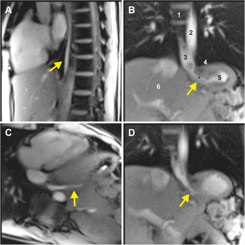
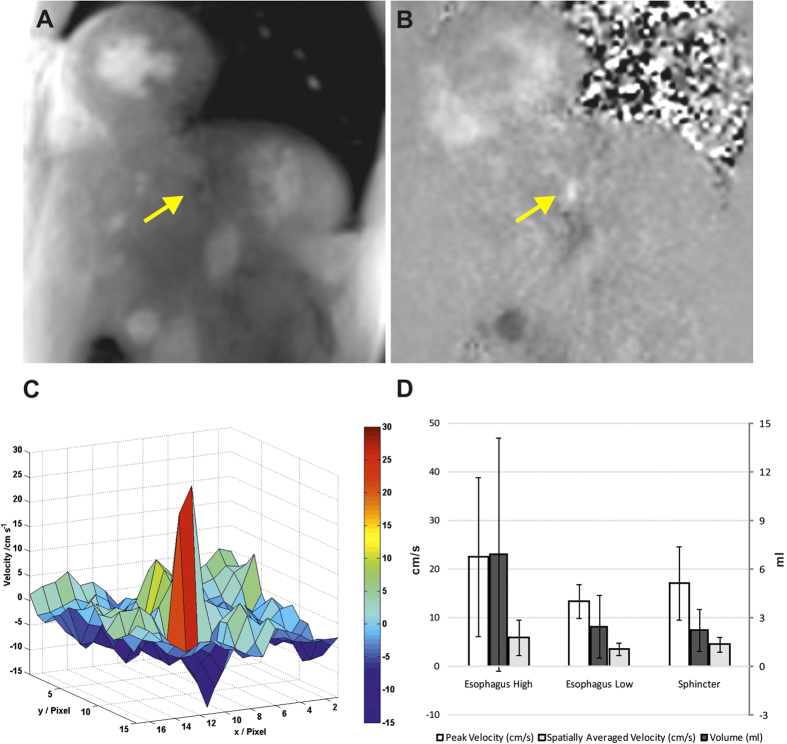
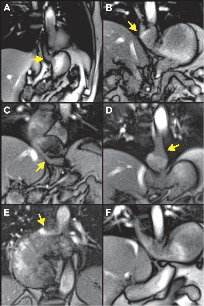
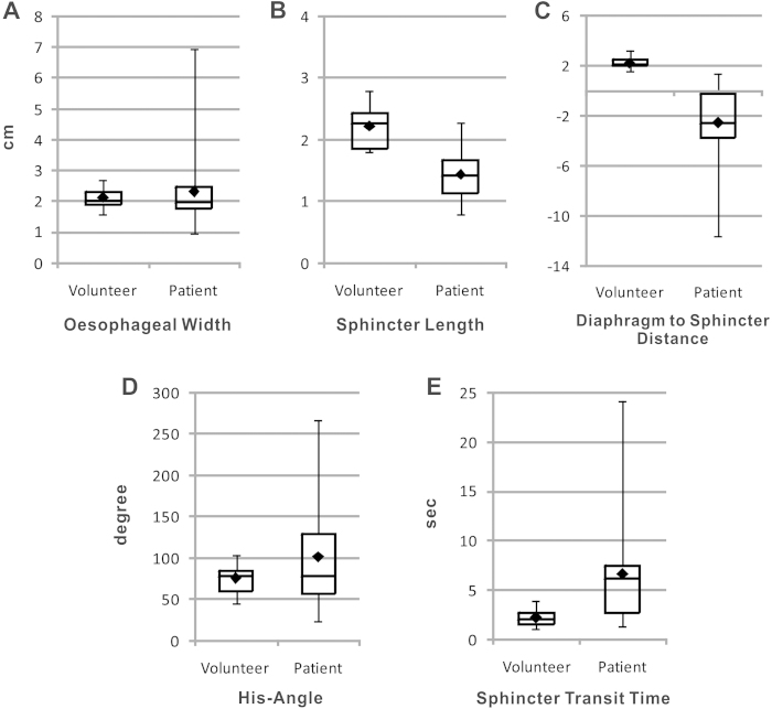
A small angle (His angle) between the oesophagus and the fundus of the stomach is considered to act as flap valve and anti-reflux barrier. A wide angle results in dysfunction of the oesophagogastric junction and subsequently in gastroesophageal reflux disease (GERD). Here, we used real-time magnetic resonance imaging (MRI) at 50 ms resolution (20 frames per second) in 12 volunteers and 12 patients with GERD to assess transport of pineapple juice through the oesophagogastric junction and reflux during Valsalva. We found that the intra-abdominal part of the oesophagus was bended towards the left side resulting in an angle of 75.3 ± 17.4, which was significantly larger during Valsava (P = 0.017). Reflux and several underlying pathologies were detected in 11 out of 12 patients. Our data visualize oesophagogastric junction physiology and disprove the flap valve hypothesis. Further, non-invasive real-time MRI has considerable potential for the diagnosis of causative pathologies leading to GERD.


Conflict of interest statement

S.Z. and J.F. hold a patent on the image acquisition and reconstruction technique used here. None of the other authors have conflicts of interest to declare.

Figures

Figure 1
Figure 1. Real-time MRI of normal gastroesophageal swallowing in a 26-year-old healthy subject.
Individual images (50 ms acquisition time, 2 × 2 mm2 resolution, 8 mm section thickness) were selected from corresponding movies (S3 to S6 in Supplementary Appendix) in (A) sagittal, (B) coronal oblique, (C) transversal oblique, and (D) coronal double-oblique planes along the directions of bolus transport (arrows). 1: spinal cord, 2: descending aorta, 3: lower oesophagus, 4: diaphragm, 5: stomach, 6: liver.
Figure 2
Figure 2. Real-time flow MRI of normal bolus transport through the lower oesophageal sphincter perpendicular to the imaging plane (same subject as in Fig. 1).
(A) Magnitude image and (B) phase-contrast flow map (6 mm section thickness, velocity sensitivity 60 cm s−1) selected from corresponding real-time flow MRI movies (S7 and S8 in Supplementary Appendix) and (C) corresponding color-coded two-dimensional velocity distribution. Arrows indicate bolus transport through the lower oesophageal sphincter into the stomach. (D) Mean values averaged across 12 healthy subjects for peak velocities and mean velocities spatially averaged over the bolus transport lumen (in cm s−1) as well as for flow volumes (in ml) at three different levels along the oesophagus.
Figure 3
Figure 3
Real-time MRI of gastroesophageal swallowing in patients with (A) reflux, (B) hiatal hernia, (C) achalasia, (D) telescoping oesophagus after fundoplication, (E) thoracic stomach, and (F) functional heartburn without reflux evidence. Individual images (50 ms acquisition time, 2 × 2 mm2 resolution, 8 mm section thickness) were selected from corresponding movies (S11 to S17 in Supplementary Appendix). Arrows indicate (A,B) bolus regurgitation from the stomach into the oesophagus during Valsalva maneuver and (CF) bolus transport from oesophagus into the stomach (CF).
Figure 4
Figure 4. Comparison of clinical real-time MRI findings in patients (n = 12) and healthy volunteers (n = 12).
Negative values of the diaphragm-to-sphincter distance indicate that the diaphragm is below the sphincter.

References

    1. Dent J., El-Serag H. B., Wallander M. A. & Johansson S. Epidemiology of gastro-oesophageal reflux disease: a systematic review. Gut 54, 710–717 (2005). - PMC - PubMed
    1. Orr W. C. Review article: sleep-related gastro-oesophageal reflux as a distinct clinical entity. Aliment Pharmacol Ther 31, 47–56 (2010). - PubMed
    1. Galmiche J. P. et al. Functional esophageal disorders. Gastroenterology 130, 1459–1465 (2006). - PubMed
    1. Bredenoord A. J., Weusten B. L., Curvers W. L., Timmer R. & Smout A. J. Determinants of perception of heartburn and regurgitation. Gut 55, 313–318 (2006). - PMC - PubMed
    1. Hill L. D. et al. The gastroesophageal flap valve: in vitro and in vivo observations. Gastrointest Endosc 44, 541–547 (1996). - PubMed

Publication types