Review
. 2015 Aug;173(2):370-8.
doi: 10.1111/bjd.13954. Epub 2015 Jul 14.

Cellular and molecular mechanisms of repair in acute and chronic wound healing

Affiliations
Review

Cellular and molecular mechanisms of repair in acute and chronic wound healing

P Martin et al. Br J Dermatol. 2015 Aug.

Abstract

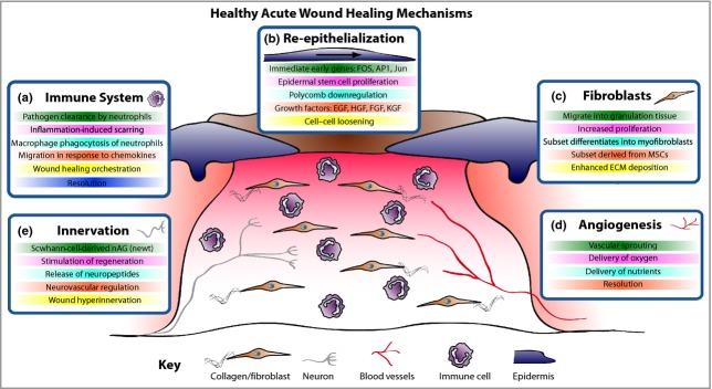
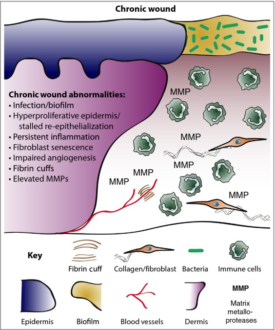
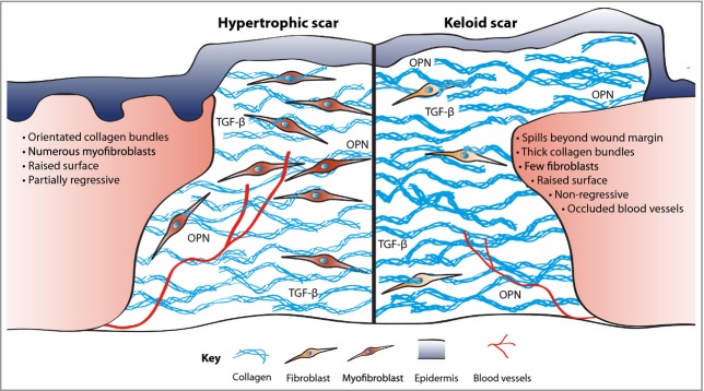
A considerable understanding of the fundamental cellular and molecular mechanisms underpinning healthy acute wound healing has been gleaned from studying various animal models, and we are now unravelling the mechanisms that lead to chronic wounds and pathological healing including fibrosis. A small cut will normally heal in days through tight orchestration of cell migration and appropriate levels of inflammation, innervation and angiogenesis. Major surgeries may take several weeks to heal and leave behind a noticeable scar. At the extreme end, chronic wounds - defined as a barrier defect that has not healed in 3 months - have become a major therapeutic challenge throughout the Western world and will only increase as our populations advance in age, and with the increasing incidence of diabetes, obesity and vascular disorders. Here we describe the clinical problems and how, through better dialogue between basic researchers and clinicians, we may extend our current knowledge to enable the development of novel potential therapeutic treatments.


Figures

Figure 1
Figure 1
Acute wound healing mechanisms. The healing of an acute wound involves coordinated cellular and molecular responses. (a) Initially immune cells migrate to the wound site and, in addition to clearing invading pathogens, in part they also orchestrate the healing process. (b) Cut epidermal edges upregulate wound-associated genes, thus enabling collective cell migration. (c) Local and blood-borne fibroblasts proliferate and migrate to form the wound granulation tissue, provide structure and signalling cues and deposit new extracellular matrix (ECM). Some fibroblasts differentiate into myofibroblasts to aid wound contraction. (d) The wound bed is perfused with oxygen and nutrients through new blood vessels derived by angiogenesis. (e) Wound healing rates exhibit a positive correlation with innervation, but hyperinnervation after wound closure could contribute to neuropathic pain. EGF, epidermal growth factor; HGF, hepatocyte growth factor; FGF, fibroblast growth factor; KGF, keratinocyte growth factor; MSC, mesenchymal stem cell; nAG, newt anterior gradient protein.
Figure 2
Figure 2
Chronic wound biology. Chronic wounds are often infected and exhibit a persistent aberrant inflammatory profile. Re-epithelialization stalls but wound keratinocytes are hyperproliferative. Granulation tissue is defective and does not nurture healing, in part due to elevated matrix metalloproteases (MMPs) and poor fibroblast infiltration. Neoangiogenesis is poor and fibrin cuffs restrict existing vessels, limiting the diffusion of oxygen through the wound, rendering the wound hypoxic.
Figure 3
Figure 3
Excessive fibrosis. Scars, formed in part as a consequence of inflammatory signals, are comprised of collagen deposited in thick orientated bundles rather than the basket-weave-like fibrils found in normal dermis. Hypertrophic scars have excessive collagen deposition, leading to a raised surface that partially resolves over time. In contrast, keloid scars have thicker collagen bundles, extend beyond the original wound margin and rarely regress. Contractile myofibroblasts are prevalent in hypertrophic scarring but all but are absent in keloid tissue. Keloids can also be characterized by occluded blood vessels. OPN, osteopontin; TGF-β, transforming growth factor-β.

References

    1. Schultz GS, White M, Mitchell R, et al. Epithelial wound healing enhanced by transforming growth factor-alpha and vaccinia growth factor. Science. 1987;235:350–2. - PubMed
    1. Grose R, Werner S. Wound-healing studies in transgenic and knockout mice. Mol Biotechnol. 2004;28:147–66. - PubMed
    1. Cooper L, Johnson C, Burslem F, Martin P. Wound healing and inflammation genes revealed by array analysis of ‘macrophageless’ PU.1 null mice. Genome Biol. 2005;6:R5. - PMC - PubMed
    1. Pedersen TX, Leethanakul C, Patel V, et al. Laser capture microdissection-based in vivo genomic profiling of wound keratinocytes identifies similarities and differences to squamous cell carcinoma. Oncogene. 2003;22:3964–76. - PubMed
    1. Paladini RD, Takahashi K, Bravo NS, Coulombe PA. Onset of re-epithelialization after skin injury correlates with a reorganization of keratin filaments in wound edge keratinocytes: defining a potential role for keratin 16. J Cell Biol. 1996;132:381–97. - PMC - PubMed

Publication types