. 2015 Oct;33(10):3006-16.
doi: 10.1002/stem.2095. Epub 2015 Jul 29.

Molecular Mechanisms Mediating Retinal Reactive Gliosis Following Bone Marrow Mesenchymal Stem Cell Transplantation

Affiliations

Molecular Mechanisms Mediating Retinal Reactive Gliosis Following Bone Marrow Mesenchymal Stem Cell Transplantation

Alessia Tassoni et al. Stem Cells. 2015 Oct.

Abstract

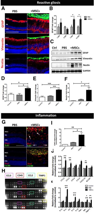
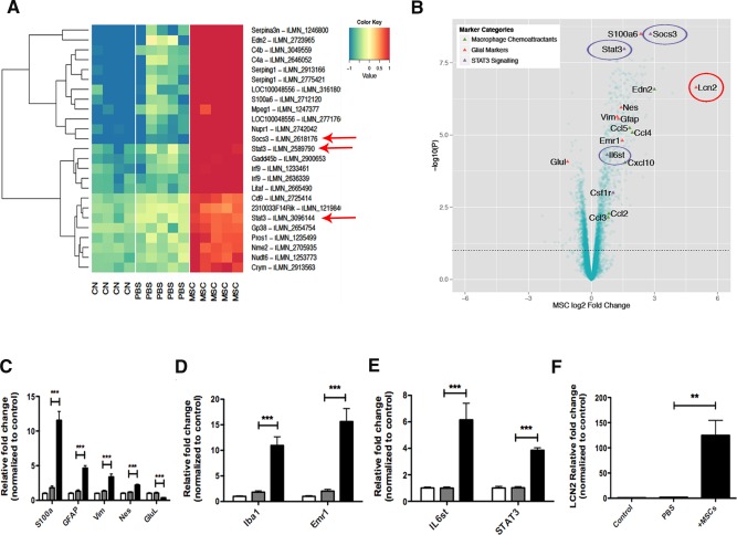
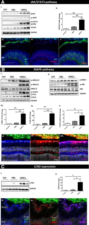
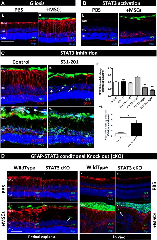
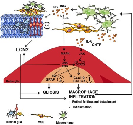
A variety of diseases lead to degeneration of retinal ganglion cells (RGCs) and their axons within the optic nerve resulting in loss of visual function. Although current therapies may delay RGC loss, they do not restore visual function or completely halt disease progression. Regenerative medicine has recently focused on stem cell therapy for both neuroprotective and regenerative purposes. However, significant problems remain to be addressed, such as the long-term impact of reactive gliosis occurring in the host retina in response to transplanted stem cells. The aim of this work was to investigate retinal glial responses to intravitreally transplanted bone marrow mesenchymal stem cells (BM-MSCs) to help identify factors able to modulate graft-induced reactive gliosis. We found in vivo that intravitreal BM-MSC transplantation is associated with gliosis-mediated retinal folding, upregulation of intermediate filaments, and recruitment of macrophages. These responses were accompanied by significant JAK/STAT3 and MAPK (ERK1/2 and JNK) cascade activation in retinal Muller glia. Lipocalin-2 (Lcn-2) was identified as a potential new indicator of graft-induced reactive gliosis. Pharmacological inhibition of STAT3 in BM-MSC cocultured retinal explants successfully reduced glial fibrillary acidic protein expression in retinal Muller glia and increased BM-MSC retinal engraftment. Inhibition of stem cell-induced reactive gliosis is critical for successful transplantation-based strategies for neuroprotection, replacement, and regeneration of the optic nerve.

Keywords: Glia; Mesenchymal stem cells; Retina; Stem cell transplantation.


Figures

Figure 1
Figure 1
MSC transplantation induces reactive gliosis and inflammation in the recipient retina. (A): Immunostaining showing upregulation of the intermediate filaments (red) GFAP, Vimentin, and Nestin following transplantation. Scale bar = 200 µm. (B): Time course of GFAP protein expression in MSC recipient retinas relative to PBS sham injected retinas (n = 4, two‐way ANOVA). (C): Western blot and its quantification (D–F) confirming gliosis at 7 days post‐transplantation. (G): Immunolabeling for Edu and F4/80 (green) with the pan microglia marker Iba1 (red) showing microglia proliferation (white arrows) and macrophage recruitment in MSC recipient retina. Scale bar = 100 µm. (H–K): Proteome profiler array and its quantification (n = 4 per group). Black, gray, and white bars represent protein expression level in MSC recipient‐, PBS sham injected‐, and naïve control retina, respectively. Error bars represent SEM, *, p < .05, **, p < .01; ***, p < .001, one‐way ANOVA test with Tukey's correction. Abbreviations: GFAP, glial fibrillary acidic protein; INL, inner nuclear layer; MSC, mesenchymal stem cell; ONL, outer nuclear layer; PBS, phosphate buffered saline; RGCL, retinal ganglion cell layer.
Figure 2
Figure 2
Microarray gene expression profiling of MSC recipient retina. (A): The top 25 probes showing the most significant changes in gene expression as ranked by ANOVA p‐value over the control (CN), sham (PBS), and MSC‐treated (MSC) groups. The expression levels of each probe across the three treatment groups are mean‐centered and are shown alongside the gene they map to. Red means high expression and blue means low expression compared to the average expression across the samples. Components of the STAT3 signaling are highlighted (red arrows). (B): Volcano plot showing key marker genes in retinal samples receiving MSC transplantation compared to PBS injected control groups. Key genes known to be markers of macrophage chemoattractant (green triangles), glial cells (red triangles), and STAT3 signaling (purple triangles) are highlighted. (C–F): Validation of the microarray data by qPCR. Error bars represent SEM, **, p < .01; ***, p < .001, one‐way ANOVA test with Tukey's correction. Abbreviations: CN, control; GFAP, glial fibrillary acidic protein; MSC, mesenchymal stem cell; PBS, phosphate buffered saline.
Figure 3
Figure 3
MSC transplantation results in LCN2 production and STAT3 and ERK activation in retinal Muller glia. Immunostaining and Western blot confirming activation of (A) JAK STAT cascade, (B) MAPK cascade, and (C) LCN2 in retinal Muller glia following MSC transplantation. Error bars represent SEM, *, p < .05; **, p < .01; ***, p < .001, one‐way ANOVA test with Tukey's correction. Abbreviations: GFAP, glial fibrillary acidic protein; GFP, green fluorescent protein; INL, inner nuclear layer; MSC, mesenchymal stem cell; ONL, outer nuclear layer; PBS, phosphate buffered saline; RGCL, retinal ganglion cell layer.
Figure 4
Figure 4
STAT3 as a central player in graft‐induced reactive gliosis ex vivo but not in vivo. (A, B): Immunostaining showing GFAP overexpression and STAT3 phosphorylation in retinal explants cocultured with MSC at 4 days ex vivo. (C): Immunostaining showing the effect of S31–201 in suppressing GFAP expression (i–iii), increasing MSC engraftment (ii, vi), and inhibiting STAT3 phosphorylation (iv, v). (D): Immunostaining showing that GFAP overexpression induced by donor MSCs is suppressed in GFAP‐STAT3 cKO mice ex vivo (i–iv) but not in vivo (vi–viii); n = 3 per group. Scale bar = 100 µm. Error bars represent SEM, *, p < .05; **, p < .01. Abbreviations: GFAP, glial fibrillary acidic protein; GFP, green fluorescent protein; INL, inner nuclear layer; MSC, mesenchymal stem cell; ONL, outer nuclear layer; PBS, phosphate buffered saline; RGCL, retinal ganglion cell layer.
Figure 5
Figure 5
Schematic diagram depicting a possible molecular mechanism orchestrating retinal gliosis and neuro‐inflammation in response to donor MSCs. Abbreviations: GFAP, glial fibrillary acidic protein; MSC, mesenchymal stem cell.

References

    1. Levin LA. Axonal loss and neuroprotection in optic neuropathies. Can J Ophthalmol 2007;42:403–408. - PubMed
    1. Johnson TV, Bull ND, Hunt DP et al. Neuroprotective effects of intravitreal mesenchymal stem cell transplantation in experimental glaucoma. Invest Ophthalmol Vis Sci 2010;51:2051–2059. - PMC - PubMed
    1. Johnson TV, DeKorver NW, Levasseur VA et al. Identification of retinal ganglion cell neuroprotection conferred by platelet‐derived growth factor through analysis of the mesenchymal stem cell secretome. Brain 2014;137:503–519. - PMC - PubMed
    1. Johnson TV, Bull ND, Martin KR. Identification of barriers to retinal engraftment of transplanted stem cells. Invest Ophthalmol Vis Sci 2010;51:960–970. - PMC - PubMed
    1. Barber AC, Hippert C, Duran Y et al. Repair of the degenerate retina by photoreceptor transplantation. Proc Natl Acad Sci USA 2013;110:354–359. - PMC - PubMed

Publication types