. 2015 Jul 13;28(1):129-40.
doi: 10.1016/j.ccell.2015.06.001.

MYC Is a Major Determinant of Mitotic Cell Fate

Affiliations

MYC Is a Major Determinant of Mitotic Cell Fate

Caroline Topham et al. Cancer Cell. .

Abstract

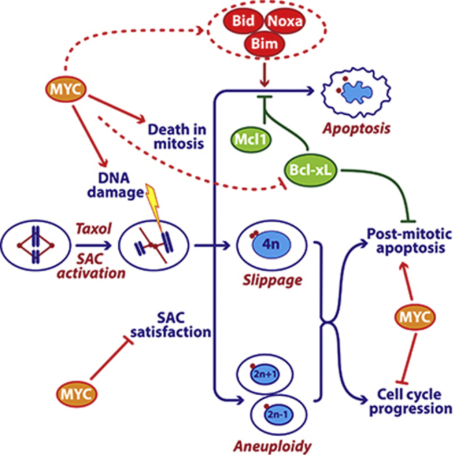
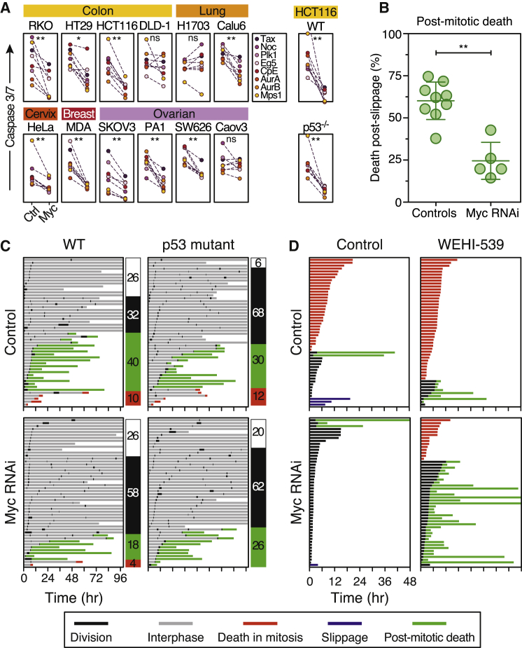
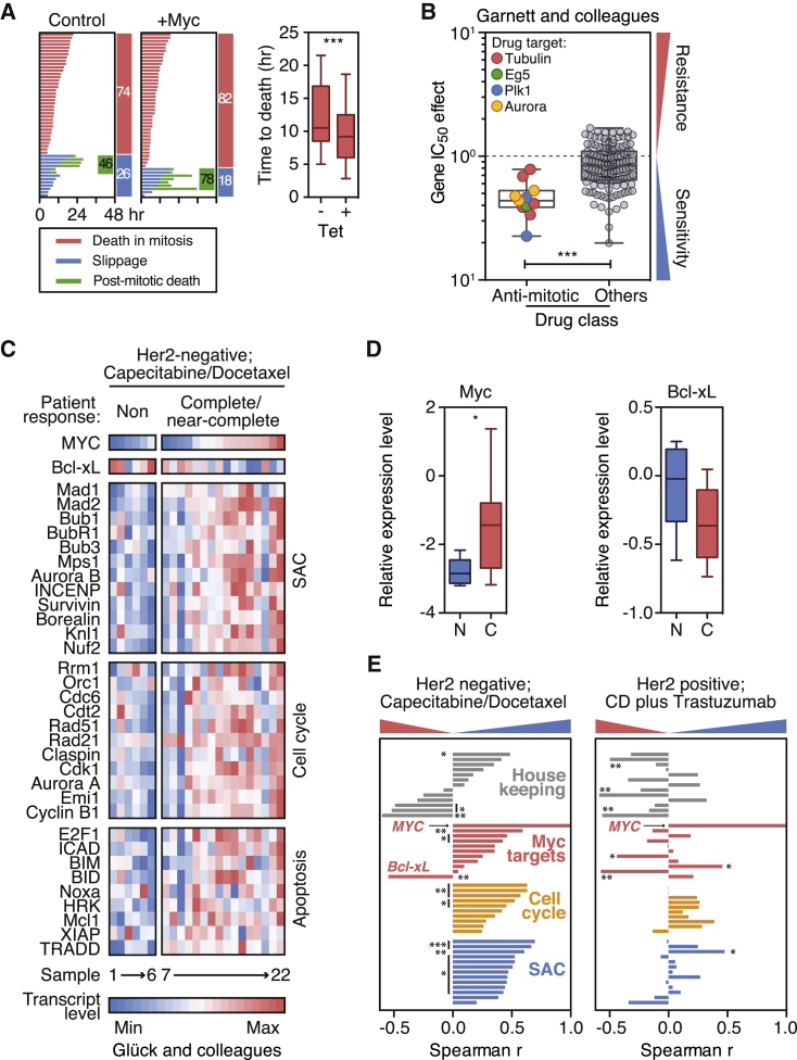
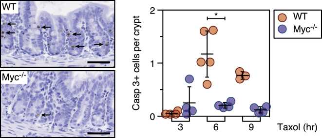
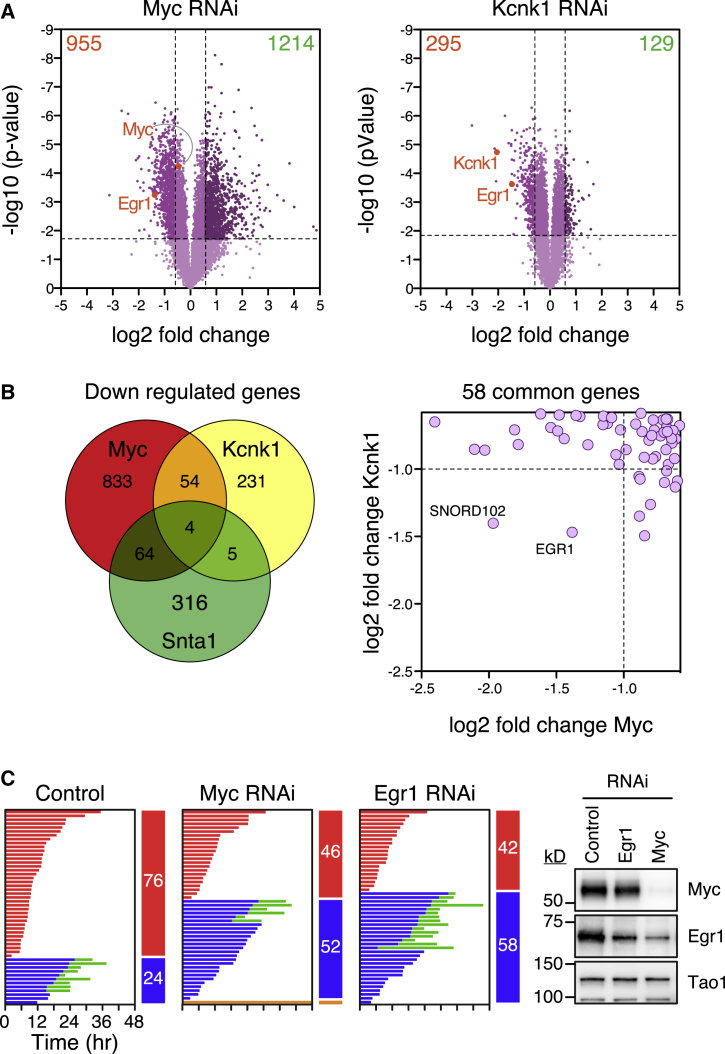
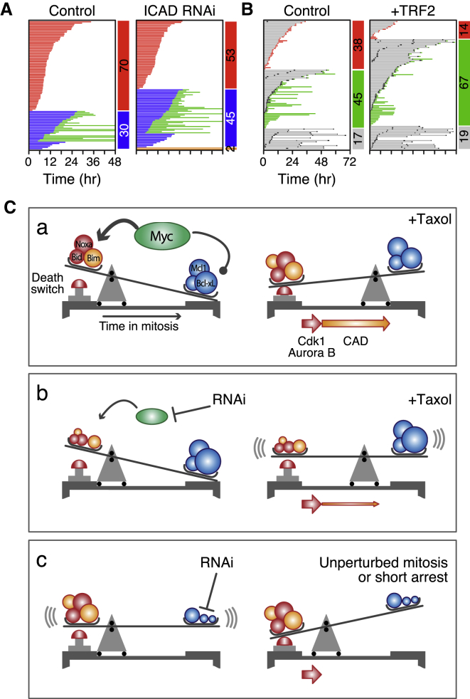
Taxol and other antimitotic agents are frontline chemotherapy agents but the mechanisms responsible for patient benefit remain unclear. Following a genome-wide siRNA screen, we identified the oncogenic transcription factor Myc as a taxol sensitizer. Using time-lapse imaging to correlate mitotic behavior with cell fate, we show that Myc sensitizes cells to mitotic blockers and agents that accelerate mitotic progression. Myc achieves this by upregulating a cluster of redundant pro-apoptotic BH3-only proteins and suppressing pro-survival Bcl-xL. Gene expression analysis of breast cancers indicates that taxane responses correlate positively with Myc and negatively with Bcl-xL. Accordingly, pharmacological inhibition of Bcl-xL restores apoptosis in Myc-deficient cells. These results open up opportunities for biomarkers and combination therapies that could enhance traditional and second-generation antimitotic agents.


Figures

None
Graphical abstract
Figure 1
Figure 1
A Genome-wide siRNA Screen for Regulators of Mitotic Cell Fate (A) Rationale for the screen based on the competing-networks model. (B) Timeline of siRNA transfection procedure. (C) Scatterplot of mitotic index at 24 hr against viability at 48 hr. See also Figure S1 and Tables S1 and S2.
Figure 2
Figure 2
Myc Is a Regulator of Mitotic Cell Fate (A) Immunoblots showing Myc inhibition. (B) Fate profiles of RKO cells exposed to 0.1 μM taxol following Myc RNAi. (C) Cell fate in nine control and five Myc RNAi populations. (D) Correlation between Myc protein levels and cell fate. (E) Fate profiles of RKO cells exposed to 0.1 μM taxol and 0.5 μM JQ1. (F) Time spent arrested in mitosis; entire population (gray), cells that die (red), and cells that slip (blue). (G) Time arrested in mitosis with lines connecting cells from the same population. ∗∗p < 0.01, ∗∗∗∗p < 0.0001. See also Figure S2.
Figure 3
Figure 3
A Cluster of Redundant BH3-Only Proteins Promote Death in Mitosis (A) Gene expression changes following Myc RNAi; y axis shows the fold change and circle sizes reflect the number of transcripts detected. Horizontal lines represent mean ± 1 SD. (B–E) Fate profiles of RKO cells exposed to 0.1 μM taxol following tet-induced overexpression of Bcl-xL (B); RNAi-mediated co-repression of Myc and Bcl-xL (C); RNAi-mediated co-repression of Bim, Bid, and Noxa (D); and tet-induced overexpression of Bim following Myc RNAi (E). See also Figure S3 and Table S3.
Figure 4
Figure 4
Myc Promotes Post-mitotic Death (A) Apoptosis induction in cell lines indicated exposed to various antimitotic agents following Myc RNAi. (B) Graph quantitating death following slippage in the presence of 0.1 μM taxol. (C) Fate profiles of wild-type and p53-deficient HCT116 cells following Myc RNAi then exposed to the Mps1 inhibitor AZ3146 (2 μM). Numbers indicate percentage of cells that undergo one division (white), multiple divisions (black), post-mitotic death (green), and DiM (red). (D) Fate profiles of RKO cells exposed to 10 nM taxol in combination with 100 nM WEHI-539. In (C), 0 hr is when imaging started.p < 0.05, ∗∗p < 0.01. See also Figure S4 and Table S4.
Figure 5
Figure 5
Overexpression of Myc Sensitizes Cancer Cells to Antimitotic Agents (A) Fate profiles and box-and-whisker plot showing time to DiM in RKO cells exposed to 0.1 μM taxol following tet-induced overexpression of Myc. (B) Gene IC50 effects for MYC comparing antimitotic agents with other drugs. (C) Heatmaps showing gene expression profiles of 22 breast tumors (six non-responders and 16 complete/near-complete responders) treated with capecitabine and docetaxel. (D) Box-and-whisker plots showing Myc and Bcl-xL expression levels in non-responsive (N) and responsive tumors (C). (E) Bar graphs showing correlations between MYC and the SAC, cell cycle, and apoptosis genes. p < 0.05, ∗∗p < 0.01, ∗∗∗p < 0.001. See also Figure S5 and Tables S5 and S6.
Figure 6
Figure 6
MYC-Deficient Crypts Are Resistant to Taxol-Induced Apoptosis Immunohistochemical staining and quantitation of cleaved caspase 3 in intestinal sections from wild-type and MYC mutant mice following 3, 6, and 9 hr exposure to taxol. Bar represents 50 μm. p < 0.05.
Figure 7
Figure 7
Egr1 Is a Regulator of Mitotic Cell Fate (A) Volcano plots showing gene expression changes following Myc and Kcnk1 RNAi. (B) Venn diagram and scatterplot showing common downregulated genes. (C) Fate profiles of RKO cells exposed to 0.1 μM taxol following Egr1 RNAi and immunoblots showing reduced Egr1 following Myc RNAi. See also Figure S6 and Table S7.
Figure 8
Figure 8
Inhibition of ICAD Enhances Slippage (A) Fate profiles of RKO cells exposed to 0.1 μM taxol following ICAD RNAi. (B) Fate profiles of RKO cells in the absence of taxol following RNAi-mediated co-repression Bcl-xL/Mcl1 plus tet-induced overexpression of TRF2. In (B), 0 hr represents when imaging started. (C) Mechanistic model, see text for details. See also Figure S7.

References

    1. A’Hern R.P., Jamal-Hanjani M., Szász A.M., Johnston S.R., Reis-Filho J.S., Roylance R., Swanton C. Taxane benefit in breast cancer—a role for grade and chromosomal stability. Nat. Rev. Clin. Oncol. 2013;10:357–364. - PubMed
    1. Bah N., Maillet L., Ryan J., Dubreil S., Gautier F., Letai A., Juin P., Barillé-Nion S. Bcl-xL controls a switch between cell death modes during mitotic arrest. Cell Death Dis. 2014;5:e1291. - PMC - PubMed
    1. Barkett M., Gilmore T.D. Control of apoptosis by Rel/NF-kappaB transcription factors. Oncogene. 1999;18:6910–6924. - PubMed
    1. Berns K., Horlings H.M., Hennessy B.T., Madiredjo M., Hijmans E.M., Beelen K., Linn S.C., Gonzalez-Angulo A.M., Stemke-Hale K., Hauptmann M. A functional genetic approach identifies the PI3K pathway as a major determinant of trastuzumab resistance in breast cancer. Cancer Cell. 2007;12:395–402. - PubMed
    1. Boone D.N., Qi Y., Li Z., Hann S.R. Egr1 mediates p53-independent c-Myc-induced apoptosis via a noncanonical ARF-dependent transcriptional mechanism. Proc. Natl. Acad. Sci. USA. 2011;108:632–637. - PMC - PubMed

Publication types

MeSH terms

Associated data