Review
. 2015 Oct;25(10):1027-42.
doi: 10.1093/glycob/cwv052. Epub 2015 Jul 14.

The multiple roles of epidermal growth factor repeat O-glycans in animal development

Affiliations
Review

The multiple roles of epidermal growth factor repeat O-glycans in animal development

Amanda R Haltom et al. Glycobiology. 2015 Oct.

Abstract

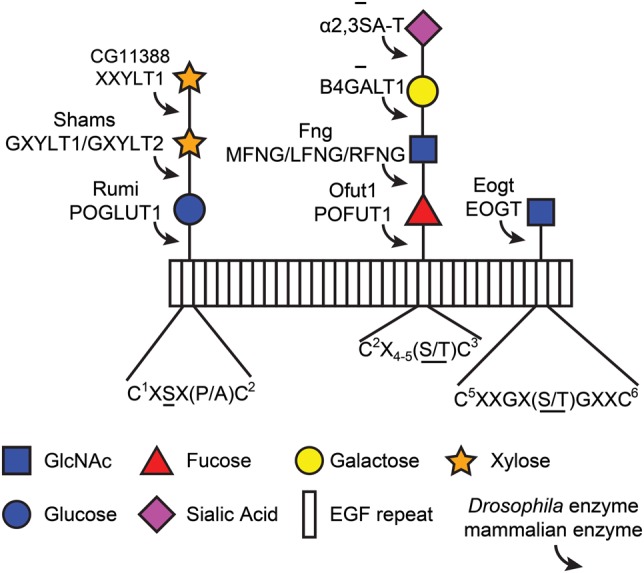
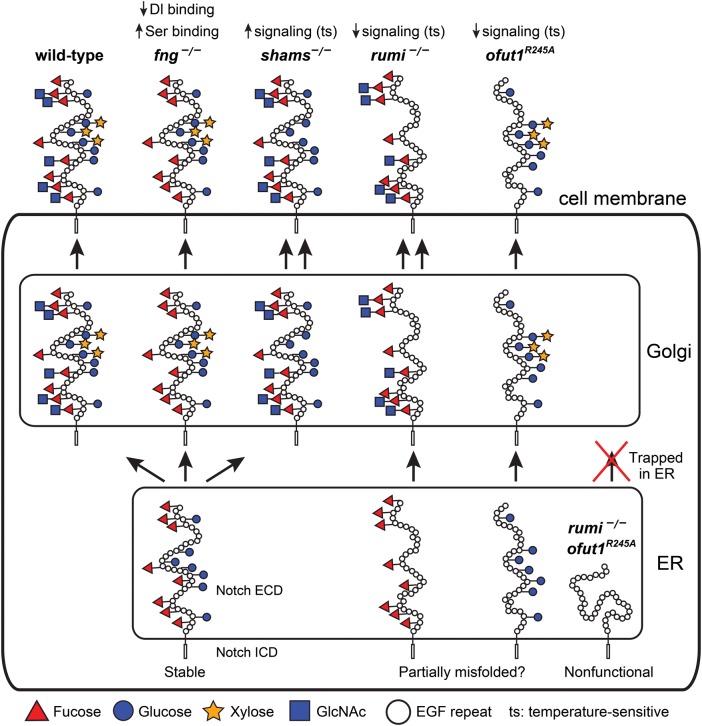
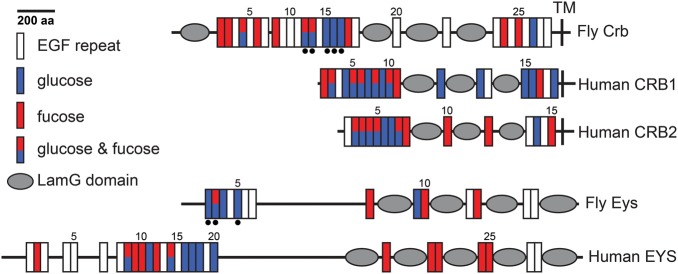
The epidermal growth factor (EGF)-like repeat is a common, evolutionarily conserved motif found in secreted proteins and the extracellular domain of transmembrane proteins. EGF repeats harbor six cysteine residues which form three disulfide bonds and help generate the three-dimensional structure of the EGF repeat. A subset of EGF repeats harbor consensus sequences for the addition of one or more specific O-glycans, which are initiated by O-glucose, O-fucose or O-N-acetylglucosamine. These glycans are relatively rare compared to mucin-type O-glycans. However, genetic experiments in model organisms and cell-based assays indicate that at least some of the glycosyltransferases involved in the addition of O-glycans to EGF repeats play important roles in animal development. These studies, combined with state-of-the-art biochemical and structural biology experiments have started to provide an in-depth picture of how these glycans regulate the function of the proteins to which they are linked. In this review, we will discuss the biological roles assigned to EGF repeat O-glycans and the corresponding glycosyltransferases. Since Notch receptors are the best studied proteins with biologically-relevant O-glycans on EGF repeats, a significant part of this review is devoted to the role of these glycans in the regulation of the Notch signaling pathway. We also discuss recently identified proteins other than Notch which depend on EGF repeat glycans to function properly. Several glycosyltransferases involved in the addition or elongation of O-glycans on EGF repeats are mutated in human diseases. Therefore, mechanistic understanding of the functional roles of these carbohydrate modifications is of interest from both basic science and translational perspectives.

Keywords: EGF repeat; Notch signaling; O-glycan; developmental biology; protein folding.


Figures

Fig. 1.
Fig. 1.
O-Linked glycans found on EGF repeats in Drosophila and mammals. Rectangles represent EGF repeats. Each modification is shown with the corresponding enzyme that adds the sugar. CG11388 is the only protein encoded by the Drosophila genome which shows a high level of homology to mammalian XXYLT1. However, its enzymatic activity remains to be verified. Addition of galactose and sialic acid to GlcNAc-fucose-O-glycans has not been observed in flies. The consensus sequence for each glycan is listed below, although the sequence for O-GlcNAc is based on a small number of confirmed sites. O-Glycans are attached to the underlined amino acid(s) in each consensus sequence. Note that a single EGF repeat can possess all three modifications (Matsuura et al. 2008). C, cysteine (the superscript numbers show the position of cysteines in the EGF repeat); X, any amino acid other than cysteine; S, serine; T, threonine; P, proline; A, alanine; Fng, Fringe; MFNG, manic fringe; LFNG, lunatic fringe; RFNG, radical fringe. This figure is available in black and white in print and in color at Glycobiology online.
Fig. 2.
Fig. 2.
Summary of the roles of O-glucose, xylose, O-fucose and GlcNAc in Drosophila Notch signaling. Schematic of the Notch protein in the ER, Golgi and at the cell membrane with its EGF repeat O-glucose and O-fucose glycans are shown in wild-type and various mutant backgrounds. The non-enzymatic chaperone function of Ofut1, which is not reported for its mammalian homologs, is not shown in this figure. Although a non-enzymatic activity has not been formally ruled out for the fly Rumi, O-glucose mutations in Notch recapitulate the rumi loss-of-function phenotypes in the context of Notch signaling. Therefore, rumi mutation is assumed to be equivalent to loss of O-glucose. For simplicity, only EGF repeats are drawn in the extracellular domain, and the intracellular domain is not drawn to scale. The folding of the extracellular domain is arbitrarily drawn. Since Drosophila Notch without O-glucose or O-fucose reaches the cell surface but shows a temperature-sensitive loss of signaling, the extracellular domain of Notch without either of these glycans is drawn as somewhat misfolded. However, other mechanisms might underlie the observed phenotypes. In the absence of both glycans, the Drosophila Notch is trapped in the ER, hence the misfolded schematic. For details, please see the text. It is important to note that the models proposed in this figure are strictly based on Drosophila studies. This figure is available in black and white in print and in color at Glycobiology online.
Fig. 3.
Fig. 3.
A model for the regulation of Eyes shut and IRS formation by Rumi. (A) A scanning electron micrograph (SEM) of the adult fly eye with its 760–800 ommatidia is shown to the left. The close-up shows a number of ommatidia, with sensory bristles decorating alternating corners of each ommatidium. To the right is a transmission electron micrograph of a single ommatidium, showing seven photoreceptor cells (PRCs). SEM images are courtesy of Jessica Leonardi. (B) Schematic drawings of a developing ommatidium. At 45% PD, the apical surfaces of photoreceptors contact one another and rhabdomere formation has not started. By 70% PD, disc-shaped rhabdomeres are formed at the apical side of photoreceptors, and secretion of Eyes shut (Eys) has separated the apical surfaces of the rhabdomeres and has formed a continuous IRS. Most of the Eys protein, which is O-glucosylated by Rumi in wild-type animals, is found in the IRS. By 100% PD, rhabdomeres have assumed their round adult morphology and are well separated by an Eys-filled IRS. (C) In rumi null eyes, a significant amount of Eys, which should lack O-glucose and is likely misfolded, remains in the PRC body. At this stage, only a fraction of Eys is secreted into the IRS, which is smaller than the wild-type IRS at the same developmental time. By 100% PD, rumi mutants do not show Eys accumulation in the PRC bodies anymore and accumulate Eys in the IRS, whose size is comparable with wild-type IRS at this stage. However, the rhabdomere attachments are not resolved, and the IRS is not continuous, suggesting that rhabdomere separation needs to occur in a critical time window during development. The ommatidium schematics are adapted from Knust (2007). Circles in PRCs depict the nuclei. Glc, glucose; ZA, zonula adherens. This figure is available in black and white in print and in color at Glycobiology online.
Fig. 4.
Fig. 4.
Distribution of O-glucose and O-fucose consensus sequences on fly and human Eyes shut and Crumbs proteins. Domain predictions were performed by using ScanProsite (http://prosite.expasy.org/). Black dots underneath EGF repeats indicate confirmed glycosylation sites (both O-glucose and O-fucose). Signal peptides at the N-terminal of the proteins are not marked. LamG, laminin G; TM, transmembrane domain. This figure is available in black and white in print and in color at Glycobiology online.

References

    1. Abd El-Aziz MM, Barragan I, O'Driscoll CA, Goodstadt L, Prigmore E, Borrego S, Mena M, Pieras JI, El-Ashry MF, Safieh LA et al. . 2008. EYS, encoding an ortholog of Drosophila spacemaker, is mutated in autosomal recessive retinitis pigmentosa. Nat Genet. 40:1285–1287. - PMC - PubMed
    1. Abd El-Aziz MM, O'Driscoll CA, Kaye RS, Barragan I, El-Ashry MF, Borrego S, Antinolo G, Pang CP, Webster AR, Bhattacharya SS. 2010. Identification of novel mutations in the ortholog of drosophila eyes shut gene (EYS) causing autosomal recessive retinitis pigmentosa. Invest Ophthalmol Vis Sci. 51:4266–4272. - PubMed
    1. Acar M, Jafar-Nejad H, Takeuchi H, Rajan A, Ibrani D, Rana NA, Pan H, Haltiwanger RS, Bellen HJ. 2008. Rumi is a CAP10 domain glycosyltransferase that modifies Notch and is required for Notch signaling. Cell. 132:247–258. - PMC - PubMed
    1. Alfaro JF, Gong CX, Monroe ME, Aldrich JT, Clauss TR, Purvine SO, Wang Z, Camp DG 2nd, Shabanowitz J, Stanley P et al. . 2012. Tandem mass spectrometry identifies many mouse brain O-GlcNAcylated proteins including EGF domain-specific O-GlcNAc transferase targets. Proc Natl Acad Sci USA. 109:7280–7285. - PMC - PubMed
    1. Andrawes MB, Xu X, Liu H, Ficarro SB, Marto JA, Aster JC, Blacklow SC. 2013. Intrinsic selectivity of Notch 1 for Delta-like 4 over Delta-like 1. J Biol Chem. 288:25477–25489. - PMC - PubMed

Publication types