Review
. 2015 Jul 14;8(385):re8.
doi: 10.1126/scisignal.aaa7540.

Reserve stem cells: Differentiated cells reprogram to fuel repair, metaplasia, and neoplasia in the adult gastrointestinal tract

Affiliations
Review

Reserve stem cells: Differentiated cells reprogram to fuel repair, metaplasia, and neoplasia in the adult gastrointestinal tract

Jason C Mills et al. Sci Signal. .

Abstract

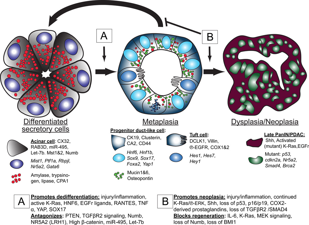
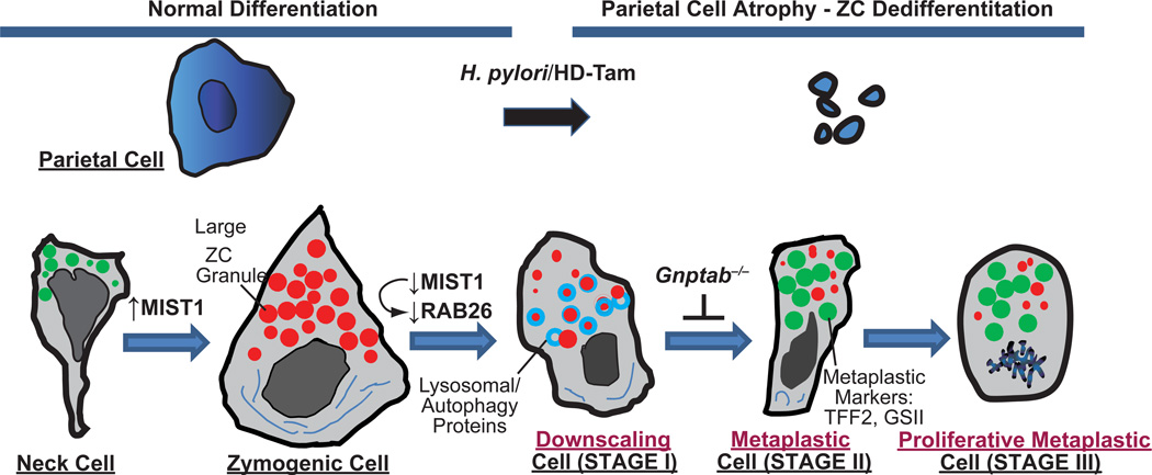
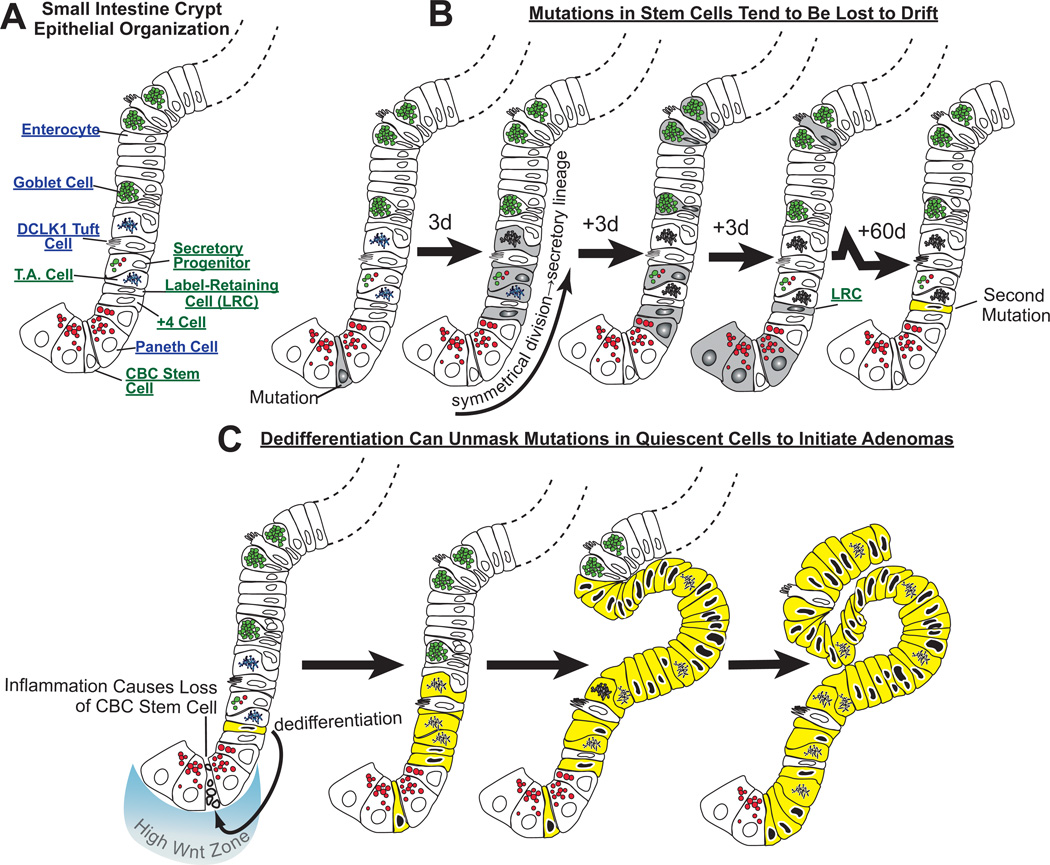
It has long been known that differentiated cells can switch fates, especially in vitro, but only recently has there been a critical mass of publications describing the mechanisms adult, postmitotic cells use in vivo to reverse their differentiation state. We propose that this sort of cellular reprogramming is a fundamental cellular process akin to apoptosis or mitosis. Because reprogramming can invoke regenerative cells from mature cells, it is critical to the long-term maintenance of tissues like the pancreas, which encounter large insults during adulthood but lack constitutively active adult stem cells to repair the damage. However, even in tissues with adult stem cells, like the stomach and intestine, reprogramming may allow mature cells to serve as reserve ("quiescent") stem cells when normal stem cells are compromised. We propose that the potential downside to reprogramming is that it increases risk for cancers that occur late in adulthood. Mature, long-lived cells may have years of exposure to mutagens. Mutations that affect the physiological function of differentiated, postmitotic cells may lead to apoptosis, but mutations in genes that govern proliferation might not be selected against. Hence, reprogramming with reentry into the cell cycle might unmask those mutations, causing an irreversible progenitor-like, proliferative state. We review recent evidence showing that reprogramming fuels irreversible metaplastic and precancerous proliferation in the stomach and pancreas. Finally, we illustrate how we think reprogrammed differentiated cells are likely candidates as cells of origin for cancers of the intestine.


Figures

Fig. 1
Fig. 1. Dedifferentiation, redifferentiation and metaplasia in the pancreas
The figure diagrams how inflammation and/or tissue damage induces terminally differentiated acinar cells with large, abundant digestive enzyme containing granules (red) to dedifferentiate to mitotically active tubuloductal structures that co-express low levels of digestive enzyme granules along with abundant granules harboring mucins and other regeneration-promoting proteins like osteopontin (green). This reprogramming process, known as acinar to ductal metaplasia, also involves increased DCLK1-positive cells akin to tuft cells of the luminal GI tract, based on gene expression and morphology. If inflammation resolves, the dedifferentiated cells can redifferentiate to regenerate normal acini (and potentially serve as stem cells to regenerate ducts and islets, though this is still an area of controversy). ADM can persist indefinitely in the presence of mutations (such as those that lead to expression of KRAS that cannot be inactivated) and/or continued inflammation. With accumulation of additional mutations, ADM can progress to pancreatic ductal adenocarcinoma (PDAC). The proteins specifically expressed at each stage of the process are delineated in the cytoplasm, nucleus, and granules. The genes involved in promoting/blocking progression to ADM (boxed “A”) and blocking redifferentiation/promoting progression to PDAC (boxed “B”) are also shown.
Fig. 2
Fig. 2. Proposed stages of reprogramming and metaplasia in the stomach
Cartoon based on our work and that of the literature proposing that reprogramming during metaplastic response to parietal cell damage in the stomach proceeds via 4 distinct stages. Zymogenic “Chief” Cells normally differentiate from a secretory progenitor, known as the mucous neck cell. Atrophy (death) of acid-secreting parietal cells (caused, e.g., by chronic infection with Helicobacter pylori or, in mouse models, by high doses of tamoxifen “HD-Tam”) causes chief cells to reprogram. We propose that the first stage of the reprogramming response is for Chief Cells to “downscale” their secretory architecture (including decreased digestive enzyme-containing granules, red, and dismantling of the elaborate rER network, dark blue) in a process involving decreased expression of the bHLH transcription factor, MIST1, which is required for maintaining apical secretory granules. Once large enzyme-containing granules are recycled, cells re-express progenitor genes (green) with much scanter digestive enzyme granules and are thus, by definition, metaplastic (called “SPEM” based on greatly increased expression of TFF2, aka Spasmolytic Polypeptide). In the third stage, cells reenter the cell cycle, at which point they may, via poorly understood processes, give rise to other metaplasias (like intestinal metaplasia in humans) or progress directly to cancer. In a parallel to pancreatic ADM, metaplastic chief cells may also regenerate other cells again via processes that are yet to be determined. Genes expressed during mucous neck, zymogenic, and metaplastic stages are delineated below, along with secreted factors at each of those stages.
Fig. 3
Fig. 3. Neoplastic growth in the intestines may occur because dedifferentiation of long-lived mature cells unmasks mutations
A) Some of the key cell lineages in the cartoon of one side of a crypt are labeled. B) Mutations occurring in a crypt base columnar (“CBC”) stem cell may be lost by drift into differentiated cells, because CBC cells divide frequently and often symmetrically to produce two daughters that differentiate. Here, a CBC cell has acquired a mutation and has undergone symmetrical division such that both daughter cells differentiate along the secretory lineage. The progeny, depicted in time intervals (e.g., “3d” ≈3 days elapsed) differentiate into goblet, Paneth, tuft and enteroendocrine (not depicted) cells. With time, most of those cells die, at which point, the mutation will be lost, were it not for the fact that some longer lived cells (e.g., Paneth cells or perhaps even longer-lived “label-retaining cells”) could potentially harbor the mutation for months. In the case depicted, the Paneth cells are also lost to normal turnover, but the even longer living LRC in this unit acquires an additional mutation over the two months following the first mutation in the stem cell. C) Again, that mutation would likely be lost as even the LRC ages and dies, unless, as is shown at the bottom, inflammation causes death of the normal, constitutively active stem cell in the crypt. Here, CBC death induces the LRC, which has accumulated an additional mutation, to dedifferentiate, become exposed to high Wnt signaling and reenter the cell cycle. The mutations in the LRC are now unmasked as the cell is exposed to the high Wnt environment and begins to proliferate. The mutations prevent the cell from differentiating beyond the transit amplifying stage, and a clone of early neoplastic cells derived from this mutant stem cell develops an adenoma. The key aspect of this model is that the normal CBC divides too rapidly to accumulate mutations, whereas differentiated cells live longer and can store mutations that may affect proliferation or differentiation but won’t affect these quiescent cells until dedifferentiation unmasks them.

References

    1. Waddington CH. The Strategy of the Genes. A Discussion of Some Aspects of Theoretical Biology. London: Geoge Allen & Unwin; 1957.
    1. Hay ED. The fine structure of blastema cells and differentiating cartilage cells in regenerating limbs of Amblystoma larvae. J Biophys Biochem Cytol. 1958;4:583–591. - PMC - PubMed
    1. Mashanov VS, Zueva OR, Garcia-Arraras JE. Expression of pluripotency factors in echinoderm regeneration. Cell Tissue Res. 2015;359:521–536. - PMC - PubMed
    1. Mosher C. Observation on evisceration and visceral regeneration in the sea-cucumber, Actinopyga agassizi Selenka. Zoologica (NY) 1956;41:17–26.
    1. Zipori D. The stem state: plasticity is essential, whereas self-renewal and hierarchy are optional. Stem Cells. 2005;23:719–726. - PubMed

Publication types

Substances

LinkOut - more resources