

A Phase I/II Study for Analytic Validation of 89Zr-J591 ImmunoPET as a Molecular Imaging Agent for Metastatic Prostate Cancer
- PMID: 26175541
- PMCID: PMC4668231
- DOI: 10.1158/1078-0432.CCR-15-0552
A Phase I/II Study for Analytic Validation of 89Zr-J591 ImmunoPET as a Molecular Imaging Agent for Metastatic Prostate Cancer
Abstract
Purpose: Standard imaging for assessing osseous metastases in advanced prostate cancer remains focused on altered bone metabolism and is inadequate for diagnostic, prognostic, or predictive purposes. We performed a first-in-human phase I/II study of (89)Zr-DFO-huJ591 ((89)Zr-J591) PET/CT immunoscintigraphy to assess performance characteristics for detecting metastases compared with conventional imaging modalities (CIM) and pathology.
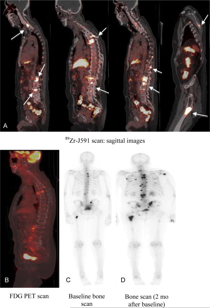
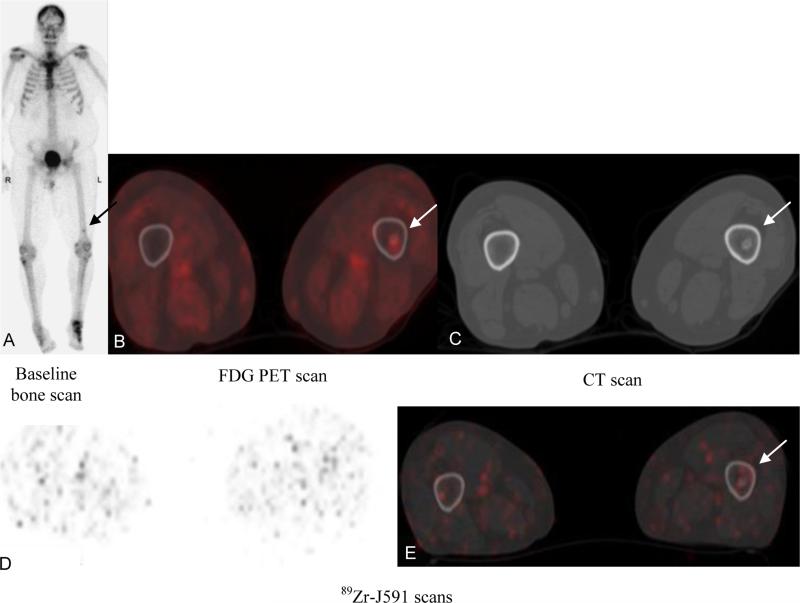
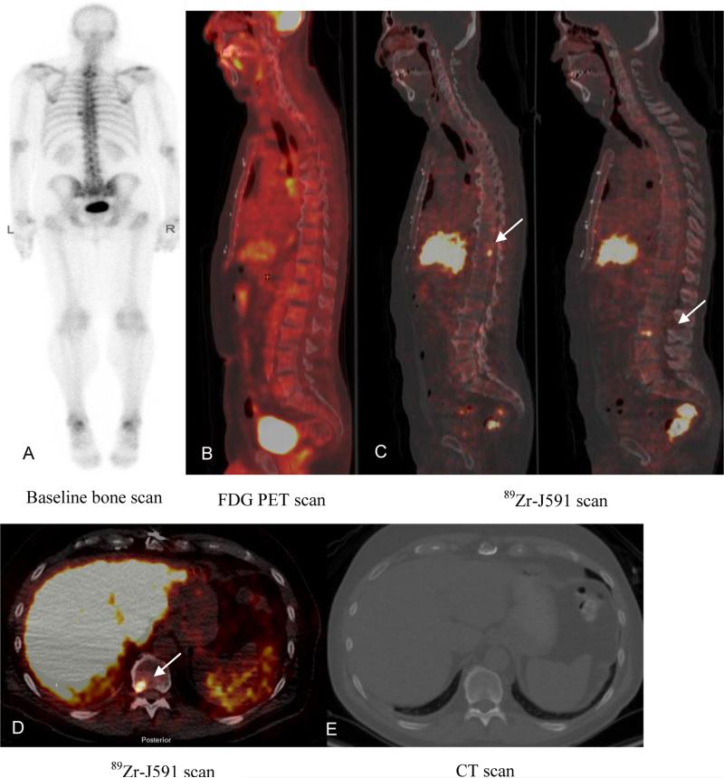
Experimental design: Fifty patients with progressive metastatic castration-resistant prostate cancers were injected with 5 mCi of (89)Zr-J591. Whole-body PET/CT scans were obtained, and images were analyzed for tumor visualization. Comparison was made to contemporaneously obtained bone scintigraphy and cross-sectional imaging on a lesion-by-lesion basis and with biopsies of metastatic sites.
Results: Median standardized uptake value for (89)Zr-J591-positive bone lesions (n = 491) was 8.9 and for soft-tissue lesions (n = 90), it was 4.8 (P < 0.00003). (89)Zr-J591 detected 491 osseous sites compared with 339 by MDP and 90 soft-tissue lesions compared with 124 by computed tomography (CT). Compared with all CIMs combined, (89)Zr-J591 detected an additional 99 osseous sites. Forty-six lesions (21 bone and 25 soft tissue) were biopsied in 34 patients; 18 of 19 (89)Zr-J591-positive osseous sites and 14 of 16 (89)Zr-J591-positive soft tissue sites were positive for prostate cancer. The overall accuracy of (89)Zr-J591 was 95.2% (20 of 21) for osseous lesions and 60% (15 of 25) for soft-tissue lesions.
Conclusions: (89)Zr-J591 imaging demonstrated superior targeting of bone lesions relative to CIMs. Targeting soft-tissue lesions was less optimal, although (89)Zr-J591 had similar accuracy as individual CIMs. This study will provide benchmark data for comparing performance of proposed prostate-specific membrane antigen (PSMA) targeting agents for prostate cancer.
©2015 American Association for Cancer Research.
Figures
References
-
- Sengoku T, Matsumura K, Usami M, Takahashi Y, Nakayama T. Diagnostic accuracy of FDG PET cancer screening in asymptomatic individuals: use of record linkage from the Osaka Cancer Registry. Int J Clin Oncol. 2014;19(6):989–97. - PubMed
-
- Minamimoto R, Uemura H, Sano F, Terao H, Nagashima Y, Yamanaka S, et al. The potential of FDG-PET/CT for detecting prostate cancer in patients with an elevated serum PSA level. Ann Nucl Med. 2011;25(1):21–7. - PubMed
Publication types
MeSH terms
Substances
Grants and funding
LinkOut - more resources
Full Text Sources
Other Literature Sources
Medical
Miscellaneous
