

Permeability Parameters Measured with Dynamic Contrast-Enhanced MRI: Correlation with the Extravasation of Evans Blue in a Rat Model of Transient Cerebral Ischemia
- PMID: 26175578
- PMCID: PMC4499543
- DOI: 10.3348/kjr.2015.16.4.791
Permeability Parameters Measured with Dynamic Contrast-Enhanced MRI: Correlation with the Extravasation of Evans Blue in a Rat Model of Transient Cerebral Ischemia
Abstract
Objective: The purpose of this study was to correlate permeability parameters measured with dynamic contrast-enhanced magnetic resonance imaging (DCE-MRI) using a clinical 3-tesla scanner with extravasation of Evans blue in a rat model with transient cerebral ischemia.
Materials and methods: Sprague-Dawley rats (n = 13) with transient middle cerebral artery occlusion were imaged using a 3-tesla MRI with an 8-channel wrist coil. DCE-MRI was performed 12 hours, 18 hours, and 36 hours after reperfusion. Permeability parameters (K(trans), ve, and vp) from DCE-MRI were calculated. Evans blue was injected after DCE-MRI and extravasation of Evans blue was correlated as a reference with the integrity of the blood-brain barrier. Correlation analysis was performed between permeability parameters and the extravasation of Evans blue.
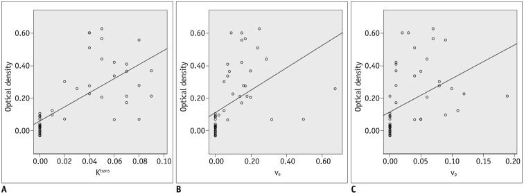
Results: All permeability parameters (K(trans), ve, and vp) showed a linear correlation with extravasation of Evans blue. Among them, K(trans) showed highest values of both the correlation coefficient and the coefficient of determination (0.687 and 0.473 respectively, p < 0.001).
Conclusion: Permeability parameters obtained by DCE-MRI at 3-T are well-correlated with Evans blue extravasation, and K(trans) shows the strongest correlation among the tested parameters.
Keywords: Animal models; Blood-brain barrier; Brain ischemia; Dynamic contrast-enhanced MRI; Kinetic modeling.
Figures


Similar articles
-
Assessment of Blood-Brain Barrier Permeability by Dynamic Contrast-Enhanced MRI in Transient Middle Cerebral Artery Occlusion Model after Localized Brain Cooling in Rats.Korean J Radiol. 2016 Sep-Oct;17(5):715-24. doi: 10.3348/kjr.2016.17.5.715. Epub 2016 Aug 23. Korean J Radiol. 2016. PMID: 27587960 Free PMC article.
-
Dynamic contrast-enhanced MRI and CT provide comparable measurement of blood-brain barrier permeability in a rodent stroke model.Magn Reson Imaging. 2015 Oct;33(8):1007-12. doi: 10.1016/j.mri.2015.06.021. Epub 2015 Jun 25. Magn Reson Imaging. 2015. PMID: 26117703
-
Neuroprotective effect of agmatine in rats with transient cerebral ischemia using MR imaging and histopathologic evaluation.Magn Reson Imaging. 2013 Sep;31(7):1174-81. doi: 10.1016/j.mri.2013.03.026. Epub 2013 May 1. Magn Reson Imaging. 2013. PMID: 23642800
-
Magnetic resonance imaging of blood-brain barrier permeability in ischemic stroke using diffusion-weighted arterial spin labeling in rats.J Cereb Blood Flow Metab. 2017 Aug;37(8):2706-2715. doi: 10.1177/0271678X16673385. Epub 2016 Jan 1. J Cereb Blood Flow Metab. 2017. PMID: 27742887 Free PMC article.
-
Markers for blood-brain barrier integrity: how appropriate is Evans blue in the twenty-first century and what are the alternatives?Front Neurosci. 2015 Oct 29;9:385. doi: 10.3389/fnins.2015.00385. eCollection 2015. Front Neurosci. 2015. PMID: 26578854 Free PMC article. Review.
Cited by
-
Alterations in the Temporal Variation and Spatial Distribution of Blood-Brain Barrier Permeability Following Electromagnetic Pulse Radiation: A Study Based on Dynamic Contrast-Enhanced MRI.Brain Sci. 2025 May 27;15(6):577. doi: 10.3390/brainsci15060577. Brain Sci. 2025. PMID: 40563749 Free PMC article.
-
Low-Dose Evans Blue Dye for Near-Infrared Fluorescence Imaging in Photothrombotic Stroke Model.Int J Med Sci. 2018 Apr 27;15(7):696-702. doi: 10.7150/ijms.24257. eCollection 2018. Int J Med Sci. 2018. PMID: 29910674 Free PMC article.
-
Emerging Techniques in Brain Tumor Imaging: What Radiologists Need to Know.Korean J Radiol. 2016 Sep-Oct;17(5):598-619. doi: 10.3348/kjr.2016.17.5.598. Epub 2016 Aug 23. Korean J Radiol. 2016. PMID: 27587949 Free PMC article. Review.
-
A Novel Histological Technique to Assess Severity of Traumatic Brain Injury in Rodents: Comparisons to Neuroimaging and Neurological Outcomes.Front Neurosci. 2021 Oct 13;15:733115. doi: 10.3389/fnins.2021.733115. eCollection 2021. Front Neurosci. 2021. PMID: 34720861 Free PMC article.
-
Knockdown of NADPH oxidase 4 reduces mitochondrial oxidative stress and neuronal pyroptosis following intracerebral hemorrhage.Neural Regen Res. 2023 Aug;18(8):1734-1742. doi: 10.4103/1673-5374.360249. Neural Regen Res. 2023. PMID: 36751799 Free PMC article.
References
-
- The National Institute of Neurological Disorders and Stroke rt-PA Stroke Study Group. Tissue plasminogen activator for acute ischemic stroke. N Engl J Med. 1995;333:1581–1587. - PubMed
-
- Lansberg MG, Albers GW, Wijman CA. Symptomatic intracerebral hemorrhage following thrombolytic therapy for acute ischemic stroke: a review of the risk factors. Cerebrovasc Dis. 2007;24:1–10. - PubMed
-
- Tanne D, Kasner SE, Demchuk AM, Koren-Morag N, Hanson S, Grond M, et al. Markers of increased risk of intracerebral hemorrhage after intravenous recombinant tissue plasminogen activator therapy for acute ischemic stroke in clinical practice: the Multicenter rt-PA Stroke Survey. Circulation. 2002;105:1679–1685. - PubMed
-
- The NINDS t-PA Stroke Study Group. Intracerebral hemorrhage after intravenous t-PA therapy for ischemic stroke. Stroke. 1997;28:2109–2118. - PubMed
-
- Barber PA, Demchuk AM, Zhang J, Buchan AM. Validity and reliability of a quantitative computed tomography score in predicting outcome of hyperacute stroke before thrombolytic therapy. ASPECTS Study Group. Alberta Stroke Programme Early CT Score. Lancet. 2000;355:1670–1674. - PubMed
MeSH terms
Substances
LinkOut - more resources
Full Text Sources
Other Literature Sources
Medical
