

doi: 10.3348/kjr.2015.16.4.853.
Epub 2015 Jul 1.
Unusual Malignant Solid Neoplasms of the Kidney: Cross-Sectional Imaging Findings
Affiliations
- PMID: 26175585
- PMCID: PMC4499550
- DOI: 10.3348/kjr.2015.16.4.853
Item in Clipboard
Unusual Malignant Solid Neoplasms of the Kidney: Cross-Sectional Imaging Findings
Korean J Radiol.
2015 Jul-Aug.
Abstract
Malignant kidney neoplasms are the most frequently encountered solid kidney masses. Although renal cell carcinoma is the major renal malignancy, other solid malignant renal masses should be considered in the differential diagnosis of solid renal masses that do not contain a macroscopic fatty component. In this pictorial essay, we present the imaging findings of a primitive neuroectodermal tumor, primary liposarcoma of the kidney, primary neuroendocrine tumor, leiomyosarcoma, synovial sarcoma, malignant fibrous histiocytoma, sclerosing fibrosarcoma and renal metastasis of osteosarcoma.
Keywords: Computed tomography; Kidney; Magnetic resonance imaging; Malignant tumors.
Figures

Axial (A) and coronal (B) post-contrast computed tomography images demonstrate large masses (arrows) arising from left kidney. Kidney parenchyma is almost completely replaced by mass. Central hypodense area (arrowheads) represents necrosis.

Axial post-contrast computed tomography image of 67-year-old male with surgically proven liposarcoma of kidney shows non-specific contrast enhancement and well circumscribed mass in upper pole of left kidney (arrow) with no associated macroscopic fat.

Axial post-contrast computed tomography image of 52-year-old male demonstrates contrast-enhanced lesion of left kidney (arrows) that was malignant neuroendocrine tumor. Punctate focus of calcification at medial edge of lesion (arrowhead) is renal stone in adjacent calyx.

A. Axial post-contrast computed tomography image demonstrates hypoenhancing solid mass with well-defined lobulated margins (arrow). Axial (B) and coronal (C) T2-weighted magnetic resonance image (MRI) reveals low signal intense left renal mass (arrows). D. Renal mass (arrow) appears with low enhancement on contrast-enhanced fat saturated T1-weighted MRI.

Axial T1- (A) and T2-weighted (B) magnetic resonance image (MRI) demonstrate right renal mass with low signal-intensity and solid (arrows) and high signal-intensity cystic (arrowheads) components. High signal intensity in cystic portion of mass represents hemorrhagic or protein rich fluid. C. Coronal T2-weighted MRI reveals right renal mass (arrow) with peripheral cystic portion. D. Axial fat-saturated T1-weighted MRI demonstrates enhancement of solid portion (arrow) of renal synovial sarcoma.

A. Unenhanced axial computed tomography (CT) image demonstrates well-defined solid mass (arrow) arising from left renal capsule. B. Contrast-enhanced axial CT image reveals heterogeneous enhancement in mass (arrow).

Axial (A) and coronal (B) contrast-enhanced computed tomography scans show low-attenuated well-defined mass (arrow) arising from left kidney.
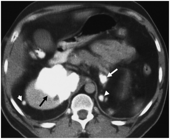
Axial post-contrast computed tomography image of 19-year-old boy with metastatic osteosarcoma of lower extremity demonstrates large calcified metastatic deposit in right kidney (black arrow). Note calcified left adrenal (white arrow) and bilateral perinephric space (arrowheads) metastases.
Similar articles
-
Pediatric and adult primary sarcomas of the kidney: a cross-sectional imaging review.Acta Radiol. 2011 May 1;52(4):448-57. doi: 10.1258/ar.2011.100376. Epub 2011 Mar 17. Acta Radiol. 2011. PMID: 21498303
-
Renal sarcoma and sarcomatoid renal cell carcinoma: CT and angiographic features.Radiology. 1987 Feb;162(2):353-7. doi: 10.1148/radiology.162.2.3797647. Radiology. 1987. PMID: 3797647
-
Primary intrathoracic malignant mesenchymal tumors: pictorial essay.J Thorac Imaging. 1994 Summer;9(3):148-55. doi: 10.1097/00005382-199422000-00006. J Thorac Imaging. 1994. PMID: 8083929
-
Synovial sarcoma of the kidney: a report of 4 cases with pathologic appraisal and differential diagnostic review.Anal Quant Cytol Histol. 2010 Aug;32(4):239-45. Anal Quant Cytol Histol. 2010. PMID: 21434526 Review.
-
CT and MRI of small renal masses.Br J Radiol. 2018 Jul;91(1087):20180131. doi: 10.1259/bjr.20180131. Epub 2018 May 10. Br J Radiol. 2018. PMID: 29668296 Free PMC article. Review.
Cited by
-
Renal Tumors of Childhood: Radiologic-Pathologic Correlation Part 2. The 2nd Decade: From the Radiologic Pathology Archives.Radiographics. 2017 Sep-Oct;37(5):1538-1558. doi: 10.1148/rg.2017160189. Radiographics. 2017. PMID: 28898190 Free PMC article. Review.
-
Primary Renal Leiomyosarcoma: A Case Report of a Rare and Aggressive Neoplasm.Cureus. 2025 Jul 1;17(7):e87099. doi: 10.7759/cureus.87099. eCollection 2025 Jul. Cureus. 2025. PMID: 40755625 Free PMC article.
-
Comparison of Biexponential and Monoexponential Model of Diffusion-Weighted Imaging for Distinguishing between Common Renal Cell Carcinoma and Fat Poor Angiomyolipoma.Korean J Radiol. 2016 Nov-Dec;17(6):853-863. doi: 10.3348/kjr.2016.17.6.853. Epub 2016 Oct 31. Korean J Radiol. 2016. PMID: 27833401 Free PMC article.
-
Incidentally detected renal leiomyosarcoma with inferior vena cava tumor thrombus: a case report with review of the literature.Front Surg. 2025 Aug 21;12:1640444. doi: 10.3389/fsurg.2025.1640444. eCollection 2025. Front Surg. 2025. PMID: 40919118 Free PMC article.
-
Neuroendocrine Neoplasms of the Female Genitourinary Tract: A Comprehensive Overview.Cancers (Basel). 2022 Jun 30;14(13):3218. doi: 10.3390/cancers14133218. Cancers (Basel). 2022. PMID: 35804996 Free PMC article. Review.
References
-
- Lee H, Cho JY, Kim SH, Jung DC, Kim JK, Choi HJ. Imaging findings of primitive neuroectodermal tumors of the kidney. J Comput Assist Tomogr. 2009;33:882–886. - PubMed
-
- Pickhardt PJ, Lonergan GJ, Davis CJ, Jr, Kashitani N, Wagner BJ. From the archives of the AFIP. Infiltrative renal lesions: radiologic-pathologic correlation. Armed Forces Institute of Pathology. Radiographics. 2000;20:215–243. - PubMed
-
- Dotan ZA, Tal R, Golijanin D, Snyder ME, Antonescu C, Brennan MF, et al. Adult genitourinary sarcoma: the 25-year Memorial Sloan-Kettering experience. J Urol. 2006;176:2033–2038. discussion 2038-2039. - PubMed
-
- Novick AC, Campbell SC. Renal tumors. In: Walsh PC, Retik AB, Vaughan ED, Wein AJ, editors. Campbell's urology. 8th ed. Philadelphia: Saunders; 2002. pp. 2673–2731.
MeSH terms
LinkOut - more resources
Full Text Sources
Other Literature Sources
Medical
