

Review
doi: 10.3348/kjr.2015.16.4.947.
Epub 2015 Jul 1.
Sclerosing Pneumocytoma with a Wax-and-Wane Pattern of Growth: A Case Report on Computed Tomography and Magnetic Resonance Imaging Findings and a Literature Review
Affiliations
- PMID: 26175598
- PMCID: PMC4499563
- DOI: 10.3348/kjr.2015.16.4.947
Item in Clipboard
Review
Sclerosing Pneumocytoma with a Wax-and-Wane Pattern of Growth: A Case Report on Computed Tomography and Magnetic Resonance Imaging Findings and a Literature Review
Korean J Radiol.
2015 Jul-Aug.
Abstract
Sclerosing pneumocytoma (SP) of the lung is a rare benign neoplasm. Here, we describe an unusual presentation of SP with a wax-and-wane pattern of growth in a 47-year-old woman. Tumor diameter decreased over a 3-year follow-up period and then increased on serial follow-up computed tomography scans. The mass showed high signal intensity on both T1- and T2-weighted chest magnetic resonance imaging (MRI) and early enhancement with a plateau on dynamic MRI. We speculate that intratumoral bleeding and resorption processes accounted for the changes in tumor size.
Keywords: Magnetic resonance imaging; Multidetector computed tomography; Pulmonary sclerosing hemangioma.
Figures
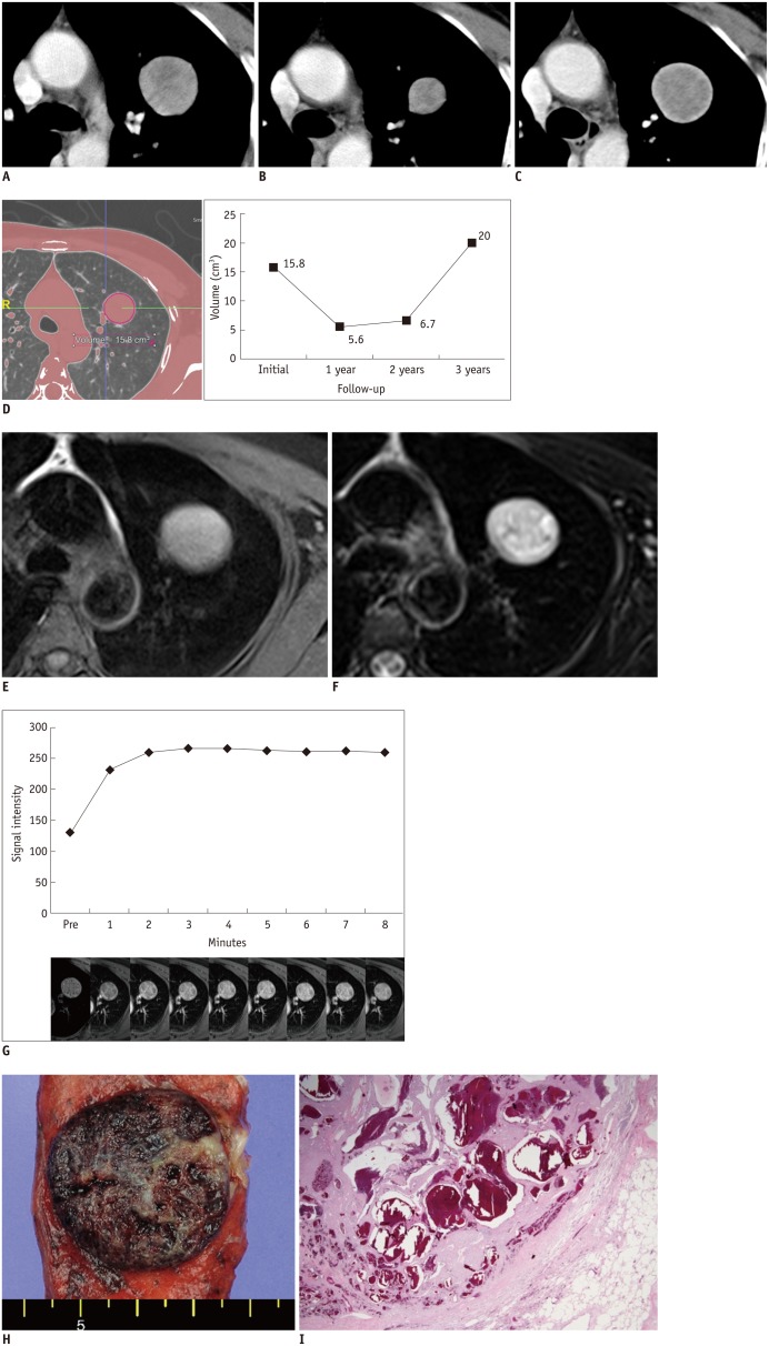
A-C. Initial (A) and 1-year (B) and 3-year follow-up (C) contrast-enhanced computed tomography (CT) scans show heterogeneously enhancing mass in left upper lobe. Mass decreases in diameter from 3.1 to 2.1 cm and then marked increase to 3.4 cm on serial CT scans. D. Serial volume graph chart obtained from three-dimensional CT data using automated segmentation technique during follow-up shows wax-and-wane pattern. E, F. Mass shows iso- to higher signal intensity (SI) than that of muscle on T1-weighted magnetic resonance (MR) image (E) and heterogeneously high SI on fat-saturated T2-weighted image (F). G. Dynamic contrast-enhanced MR images and corresponding graph of SI versus time show early enhancement without peak point and subsequent plateau pattern. H. Gross findings show well-demarcated, solid mass with fibrous matrix and areas of hemorrhage. I. Well-demarcated mass with small cystic spaces filled with blood was observed on microscopic examination (hematoxylin and eosin staining, × 12.5).
Similar articles
-
[Sclerosing pneumocytoma: an old disease with a rare presentation].Medicina (B Aires). 2024;84(6):1249-1251. Medicina (B Aires). 2024. PMID: 39666420 Spanish.
-
Clustered pulmonary sclerosing pneumocytoma in a young man: a case report.Clin Imaging. 2014 Jul-Aug;38(4):532-535. doi: 10.1016/j.clinimag.2014.01.016. Epub 2014 Feb 7. Clin Imaging. 2014. PMID: 24667045
-
A rare tumor of the lung: pulmonary sclerosing hemangioma (pneumocytoma).Respir Med. 2013 Mar;107(3):448-50. doi: 10.1016/j.rmed.2012.12.005. Epub 2013 Jan 2. Respir Med. 2013. PMID: 23290153
-
Radiology-pathology conference: sclerosing hemangioma of the lung.Clin Imaging. 2006 Nov-Dec;30(6):409-12. doi: 10.1016/j.clinimag.2006.05.030. Clin Imaging. 2006. PMID: 17101410 Review.
-
Sclerosing pneumocytoma mixed with a typical carcinoid tumor: A case report and review of literature.Medicine (Baltimore). 2019 Feb;98(5):e14315. doi: 10.1097/MD.0000000000014315. Medicine (Baltimore). 2019. PMID: 30702609 Free PMC article. Review.
Cited by
-
A case of pulmonary sclerosing pneumocytoma in the hilar lesion.Gen Thorac Cardiovasc Surg. 2019 Sep;67(9):818-820. doi: 10.1007/s11748-018-1043-6. Epub 2018 Nov 28. Gen Thorac Cardiovasc Surg. 2019. PMID: 30488193
-
18 F-FDG PET/CT imaging: A supplementary understanding of pulmonary sclerosing pneumocytoma.Thorac Cancer. 2019 Jul;10(7):1552-1560. doi: 10.1111/1759-7714.13100. Epub 2019 May 27. Thorac Cancer. 2019. PMID: 31131992 Free PMC article.
-
Magnetic resonance imaging findings of pulmonary sclerosing pneumocytoma: a case report and literature review.Front Oncol. 2023 Sep 1;13:1158328. doi: 10.3389/fonc.2023.1158328. eCollection 2023. Front Oncol. 2023. PMID: 37727218 Free PMC article.
References
-
- Illei PB, Rosai J, Klimstra DS. Expression of thyroid transcription factor-1 and other markers in sclerosing hemangioma of the lung. Arch Pathol Lab Med. 2001;125:1335–1339. - PubMed
-
- Devouassoux-Shisheboran M, Hayashi T, Linnoila RI, Koss MN, Travis WD. A clinicopathologic study of 100 cases of pulmonary sclerosing hemangioma with immunohistochemical studies: TTF-1 is expressed in both round and surface cells, suggesting an origin from primitive respiratory epithelium. Am J Surg Pathol. 2000;24:906–916. - PubMed
-
- Fujiyoshi F, Ichinari N, Fukukura Y, Sasaki M, Hiraki Y, Nakajo M. Sclerosing hemangioma of the lung: MR findings and correlation with pathological features. J Comput Assist Tomogr. 1998;22:1006–1008. - PubMed
-
- Liebow AA, Hubbell DS. Sclerosing hemangioma (histiocytoma, xanthoma) of the lung. Cancer. 1956;9:53–75. - PubMed
Publication types
MeSH terms
LinkOut - more resources
Full Text Sources
Other Literature Sources
Medical
