Review
. 2015 Jul;78(3):180-9.
doi: 10.4046/trd.2015.78.3.180. Epub 2015 Jun 30.

The International Association for the Study of Lung Cancer Lymph Node Map: A Radiologic Atlas and Review

Affiliations
Review

The International Association for the Study of Lung Cancer Lymph Node Map: A Radiologic Atlas and Review

Jin Hwan Kim et al. Tuberc Respir Dis (Seoul). 2015 Jul.

Abstract

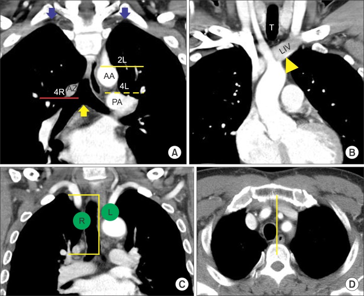
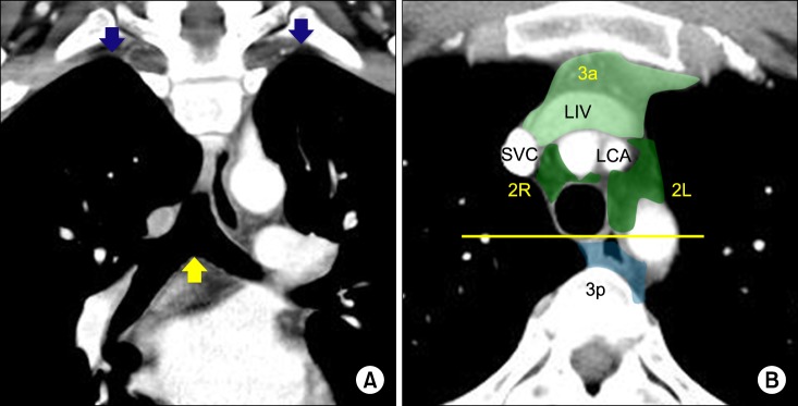
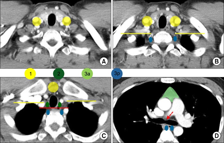
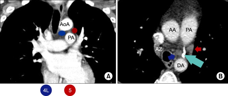
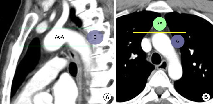
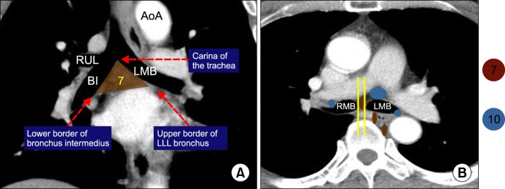
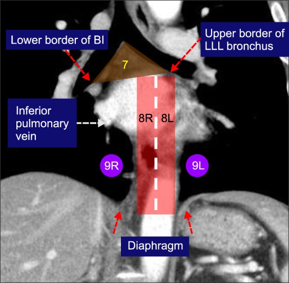
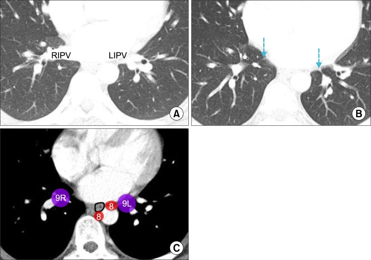
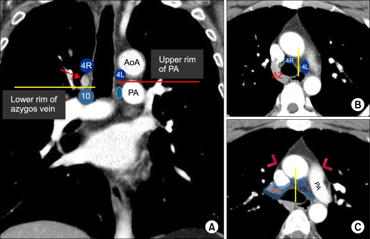
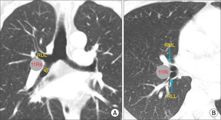
Accurate lymph node staging of lung cancer is crucial in determining optimal treatment plans and predicting patient outcome. Currently used lymph node maps have been reconciled to the internationally accepted International Association for the Study of Lung Cancer (IASLC) map published in the seventh edition of TNM classification system of malignant tumours. This article provides computed tomographic illustrations of the IASLC nodal map, to facilitate its application in day-to-day clinical practice in order to increase the appropriate classification in lung cancer staging.

Keywords: Lung Neoplasms; Lymphatic Diseasesinto; Neoplasm Staging.


Conflict of interest statement

Conflicts of Interest: No potential conflict of interest relevant to this article was reported.

Figures

Figure 1
Figure 1. Station 1. (A) The upper border of station 1 is the lower margin of the cricoid cartilage (yellow arrow). The lower border is clavicles bilaterally (C), in the midline, the upper border of the manubrium (M). (B) The border between 1R and 1L is the midline of the trachea (dashed line). C: clavicle; CR: cricoid cartilage; HY: hyoid bone; M: manubrium; TH: thyroid cartilage.
Figure 2
Figure 2. Upper zone: stations 2R, 2L, 3a, 3p, 4R, and 4L. (A, B) Upper and lower borders of stations 2, 3 and 4. The upper border of station 2R, 2L, 3a and 3p is the apex of each lung and pleural space (blue arrows), and in the midline, the upper border of the manubrium. The lower borders of the stations are different from station to station: station 2R: the intersection of caudal margin of innominate vein with the trachea (yellow triangle in B); station 2L: the superior border of the aortic arch (yellow line); station 3a and 3p, the level of carina (yellow arrow); station 4R: the lower border of the azygos vein (red line); station 4L: the upper rim of the left main pulmonary artery (yellow dashed line). (C, D) The border between 2R and 2L and between 4R and 4L moves from the midline of trachea to the left lateral border of the trachea. AA: aortic arch; AZ: azygos vein; LIV: left innominate vein; PA: left main pulmonary artery; T: trachea.
Figure 3
Figure 3. Stations 3a and 3p. (A) The upper border of station 3a and 3p is the apex of chest (blue arrows) and lower border is the level of carina (yellow arrow). (B) The anterior border of 3a is the posterior aspect of the sternum, and the posterior border is the anterior border of the superior vena cava on the right and left carotid artery on the left. Station 3p is a retrotracheal lymph node. LCA: left common carotid artery; LIV: left innominate vein; SVC: superior vena cava.
Figure 4
Figure 4. Boundary distinction among stations 1, 2 and 3. (A) Station 1 supaclavicular lymph node. (B) Inferior to the lung apex, the yellow line running horizontally from anterior margin of both lung pleura interface separates station 1 from station 3p. (C) At the suprasternal notch, the yellow line running horizontally from anterior margin of both lung pleura interface separates station 1 from station 2. The red line running along posterior wall of trachea separates station 2 from 3p. (D) Station 3a and 3p at the level of tracheal bifurcation (dashed red arrow).
Figure 5
Figure 5. Station 5. (A) The upper border of station 5 is the lower border of aortic arch (AoA), and lower border is the upper rim of the left main pulmonary artery (PA). (B) The border between station 4L and station 5 is the ligamentum arteriosum (sky-blue arrow). Station 4L nodes locate medial to the ligamentum arteriosum (blue arrow), and station 5 is the lymph node lateral to the ligamentum arteriosum (brown arrow). AA: ascending aorta; AZ: azygos vein; DA: descending aorta.
Figure 6
Figure 6. Station 6. (A) The upper border of station 6 is a line tangential to the upper border of the aortic arch and lower border is the lower border of aortic arch (green lines). (B) The anterior border of station 6 is the imaginary horizontal line extending from the anterior wall of the aortic arch (yellow line), which discriminates station 6 from station 3a. AoA: aortic arch.
Figure 7
Figure 7. Subcarinal zone: Station 7. (A) Upper and lower borders of station 7 are well identified on coronal image. (B) Station 7 nodes are noted in the space between the medial margin of both main bronchi (yellow lines) and nodes outside of the space are station 10. Station 7 extends posteriorly around the esophagus. AoA: aortic arch; BI: bronchus intermedius; LLL: left lower lobe; LMB: left main bronchus; RMB: right main bronchus; RUL: right upper lobe.
Figure 8
Figure 8. Lower zone: stations 8 and 9. The upper border of station 8 is the upper border of the left lower lobe (LLL) bronchus on the left side and the lower border of the bronchus intermedius (BI) on the right side. The upper border of the station 9 is the inferior pulmonary vein. The lower border of station 8 and 9 is the diaphragm. The border between 8R and 8L is the midline (dashed line).
Figure 9
Figure 9. Station 9 and the inferior pulmonary ligament. (A) Both inferior pulmonary veins joining the left atrium can be easily identified on computed tomography images. (B) Right and left inferior pulmonary ligaments (sky-blue dashed arrows) attach each lower lobe to the diaphragm. (C) Stations 8 and 9 nodes on axial image. Station 9 nodes locate in the inferior pulmonary ligament. Station 8 nodes are present adjacent to the esophagus (black circle). LIPV: left inferior pulmonary vein; RIPV: right inferior pulmonary vein.
Figure 10
Figure 10. Station 10. (A) The border between station 4R and 10R is the lower rim of the azygos vein (red dashed arrow). The border between station 4L and 10L is the upper rim of the left main pulmonary artery (red line). (B) Station 4R and 4L nodes at the level of azygos vein. Note the border between station 4R and 4L is the left lateral wall of the trachea (yellow line). (C) Stations 10R and 10L below the azygos vein. The pleural reflection (pink arrowhead) no longer serves as the border between station 4 and 10. The border between station 10R and 10L is the midline of tracheal bifurcation (yellow line). AoA: aortic arch; AZ: azygos vein; PA: left pulmonary artery.
Figure 11
Figure 11. Station 11R. (A) Station 11Rs is located between the right upper lobe bronchus and bronchus intermedius. (B) Station 11Ri is present between the right middle and lower lobe bronchi. Lung window setting is useful to define the bronchial details. BI: bronchus intermedius; RLL: right lower lobe; RML: right middle lobe; RUL: right upper lobe.

Similar articles

Cited by

References

    1. Ignatius Ou SH, Zell JA. The applicability of the proposed IASLC staging revisions to small cell lung cancer (SCLC) with comparison to the current UICC 6th TNM edition. J Thorac Oncol. 2009;4:300–310. - PubMed
    1. Greaves SM, Brown K, Garon EB, Garon BL. The new staging system for lung cancer: imaging and clinical implications. J Thorac Imaging. 2011;26:119–131. - PubMed
    1. Travis WD, Giroux DJ, Chansky K, Crowley J, Asamura H, Brambilla E, et al. The IASLC Lung Cancer Staging Project: proposals for the inclusion of broncho-pulmonary carcinoid tumors in the forthcoming (seventh) edition of the TNM Classification for Lung Cancer. J Thorac Oncol. 2008;3:1213–1223. - PubMed
    1. Rusch VW, Asamura H, Watanabe H, Giroux DJ, Rami-Porta R, Goldstraw P, et al. The IASLC lung cancer staging project: a proposal for a new international lymph node map in the forthcoming seventh edition of the TNM classification for lung cancer. J Thorac Oncol. 2009;4:568–577. - PubMed
    1. Naruke T, Suemasu K, Ishikawa S. Lymph node mapping and curability at various levels of metastasis in resected lung cancer. J Thorac Cardiovasc Surg. 1978;76:832–839. - PubMed