

Short stature and primary ovarian insufficiency possibly due to chromosomal position effect in a balanced X;1 translocation
- PMID: 26175800
- PMCID: PMC4501070
- DOI: 10.1186/s13039-015-0154-3
Short stature and primary ovarian insufficiency possibly due to chromosomal position effect in a balanced X;1 translocation
Abstract
Background: Primary ovarian insufficiency (POI) is defined as a primary ovarian defect characterized by absent menarche (primary amenorrhea), a decrease in the initial primordial follicle number, high follicle-stimulating hormone (FSH) levels and hypoestrogenism. Although the etiology of a majority of POI cases is not yet identified, several data suggest that POI has a strong genetic component. Conventional cytogenetic and molecular analyses have identified regions of the X chromosome that are associated with ovarian function, as well as POI candidate genes, such as FMR1 and DIAPH2. Here we describe a 10.5-year-old girl presenting with high FSH and luteinizing hormone (LH) levels, pathologic GH stimulation arginine and clonidine tests, short stature, pterygium, ovarian dysgenesis, hirsutism and POI.
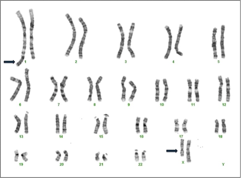
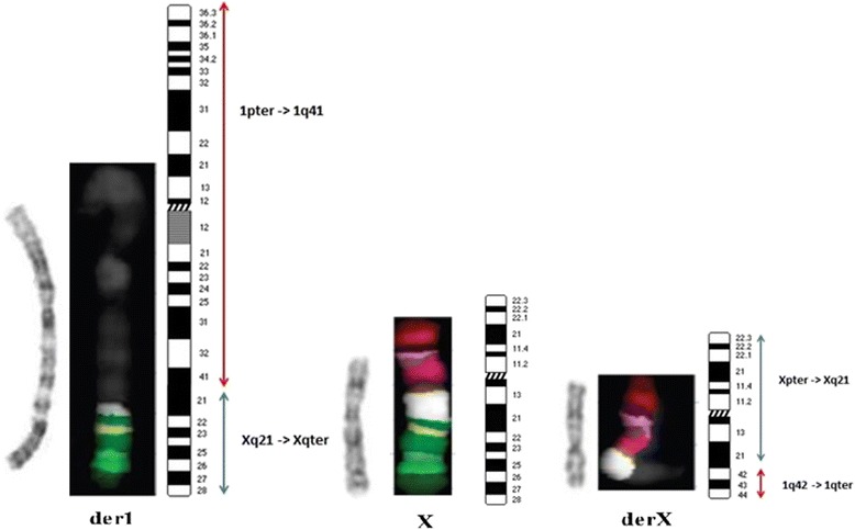
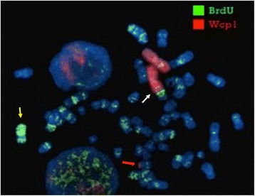
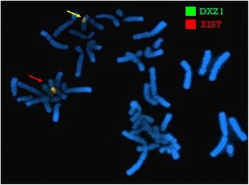
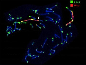
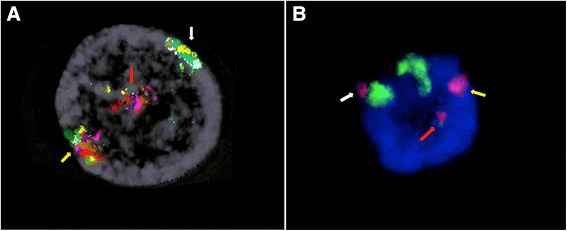
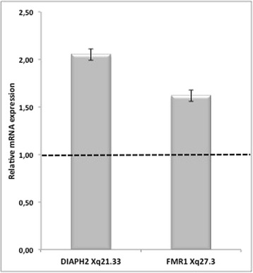
Results: Cytogenetic analysis demonstrated a balanced reciprocal translocation between the q arms of chromosomes X and 1, with breakpoints falling in Xq21 and 1q41 bands. Molecular studies did not unravel any chromosome microdeletion/microduplication, and no XIST-mediated inactivation was found on the derivative chromosome 1. Interestingly, through immunofluorescence assays, we found that part of the Xq21q22 trait, translocated to chromosome 1q41, was late replicating and therefore possibly inactivated in 30 % metaphases both in lymphocytes and skin fibroblasts, in addition to a skewed 100 % inactivation of the normal X chromosome. These findings suggest that a dysregulation of gene expression might occur in this region. Two genes mapping to the Xq translocated region, namely DIAPH2 and FMR1, were found overexpressed if compared with controls.
Conclusions: We report a case in which gonadal dysgenesis and POI are associated with over-expression of DIAPH2 gene and of FMR1 gene in wild type form. We hypothesize that this over-expression is possibly due to a phenomenon known as "chromosomal position effect", which accounts for gene expression variations depending on their localization within the nucleus. For the same effect a double mosaic inactivation of genes mapping to the Xq21-q22 region, demonstrated by immunofluorescence assays, may be the cause of a functional Xq partial monosomy leading to most Turner traits of the proband's phenotype.
Keywords: Chromosomal position effect; DIAPH2; FMR1; Primary ovarian insufficiency; Turner syndrome; X chromosome translocation; X;autosome translocation.
Figures
References
-
- Visser JA, Schipper I, Laven JS, Themmen AP. Anti-Mullerian hormone: an ovarian reserve marker in primary ovarian insufficiency. Nat Rev Endocrinol. 2012;8:331–341. - PubMed
Publication types
LinkOut - more resources
Full Text Sources
Other Literature Sources
