

Cordyceps sinensis attenuates renal fibrosis and suppresses BAG3 induction in obstructed rat kidney
- PMID: 26175854
- PMCID: PMC4494144
Cordyceps sinensis attenuates renal fibrosis and suppresses BAG3 induction in obstructed rat kidney
Abstract
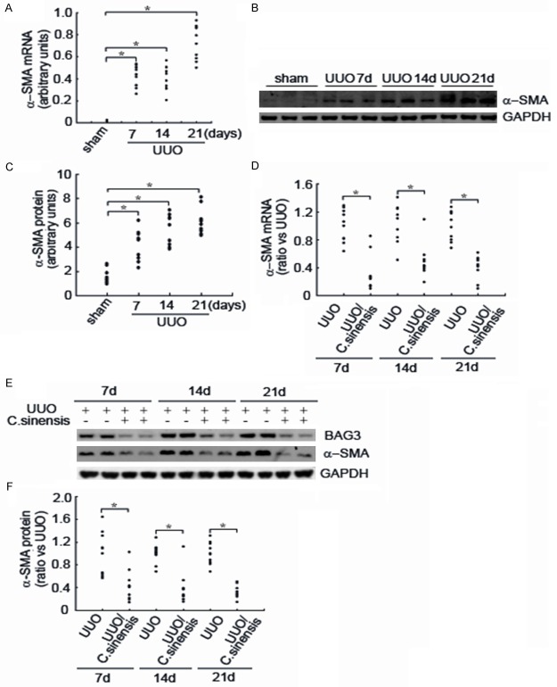
BAG3 regulates a number of cellular processes, including cell proliferation, apoptosis, adhesion and migration, and epithelial-mesenchymal transition (EMT). However, the role of BAG3 in renal tubular EMT and renal interstitial fibrosis remains elusive. This study aimed to examine the dynamic expression of BAG3 during renal fibrosis, and to investigate the efficacy of Cordyceps sinensis (C. sinensis) on renal fibrosis. A rat model of unilateral ureteral obstruction (UUO) was established, and the expression of BAG3 and α-SMA, and the efficacy of C. sinensis on renal fibrosis induced by UUO were examined. The results showed that UUO led to collagen accumulation, which was significantly suppressed by C. sinensis. UUO increased the expression of BAG3 and α-SMA, a mesenchymal marker, while UUO induced BAG3 and α-SMA expression was significantly inhibited by C. sinensis. In addition, immunohistochemical staining demonstrated that BAG3 immunoreactivity was restricted to tubular epithelium. In conclusion, BAG3 is a potential target for the prevention and/or treatment of renal fibrosis, and C. Sinensis is a promising agent for renal fibrosis.
Keywords: BAG3; Cordyceps sinensis; renal fibrosis; unilateral ureteral obstruction.
Figures


Similar articles
-
Research Progress of Cordyceps sinensis and Its Fermented Mycelium Products on Ameliorating Renal Fibrosis by Reducing Epithelial-to-Mesenchymal Transition.J Inflamm Res. 2023 Jul 7;16:2817-2830. doi: 10.2147/JIR.S413374. eCollection 2023. J Inflamm Res. 2023. PMID: 37440993 Free PMC article. Review.
-
Potential Therapeutic Strategies for Renal Fibrosis: Cordyceps and Related Products.Front Pharmacol. 2022 Jul 8;13:932172. doi: 10.3389/fphar.2022.932172. eCollection 2022. Front Pharmacol. 2022. PMID: 35873549 Free PMC article. Review.
-
Intermedin attenuates renal fibrosis by induction of heme oxygenase-1 in rats with unilateral ureteral obstruction.BMC Nephrol. 2017 Jul 11;18(1):232. doi: 10.1186/s12882-017-0659-6. BMC Nephrol. 2017. PMID: 28697727 Free PMC article.
-
Implication of Bcl-2-associated athanogene 3 in fibroblast growth factor-2-mediated epithelial-mesenchymal transition in renal epithelial cells.Exp Biol Med (Maywood). 2015 May;240(5):566-75. doi: 10.1177/1535370214558023. Epub 2014 Oct 30. Exp Biol Med (Maywood). 2015. PMID: 25361773 Free PMC article.
-
Human umbilical cord-derived mesenchymal stem cells conditioned medium attenuate interstitial fibrosis and stimulate the repair of tubular epithelial cells in an irreversible model of unilateral ureteral obstruction.Nephrology (Carlton). 2018 Aug;23(8):728-736. doi: 10.1111/nep.13099. Nephrology (Carlton). 2018. PMID: 28667820
Cited by
-
Chemical components in cultivated Cordyceps sinensis and their effects on fibrosis.Chin Herb Med. 2023 May 25;16(1):162-167. doi: 10.1016/j.chmed.2022.11.008. eCollection 2024 Jan. Chin Herb Med. 2023. PMID: 38375041 Free PMC article.
-
Use of bailing capsules (cordyceps sinensis) in the treatment of chronic kidney disease: a meta-analysis and network pharmacology.Front Pharmacol. 2024 Apr 5;15:1342831. doi: 10.3389/fphar.2024.1342831. eCollection 2024. Front Pharmacol. 2024. PMID: 38645562 Free PMC article. Review.
-
Research Progress of Cordyceps sinensis and Its Fermented Mycelium Products on Ameliorating Renal Fibrosis by Reducing Epithelial-to-Mesenchymal Transition.J Inflamm Res. 2023 Jul 7;16:2817-2830. doi: 10.2147/JIR.S413374. eCollection 2023. J Inflamm Res. 2023. PMID: 37440993 Free PMC article. Review.
-
Potential Therapeutic Strategies for Renal Fibrosis: Cordyceps and Related Products.Front Pharmacol. 2022 Jul 8;13:932172. doi: 10.3389/fphar.2022.932172. eCollection 2022. Front Pharmacol. 2022. PMID: 35873549 Free PMC article. Review.
-
Bailing capsule (Cordyceps sinensis) ameliorates renal triglyceride accumulation through the PPARα pathway in diabetic rats.Front Pharmacol. 2022 Aug 25;13:915592. doi: 10.3389/fphar.2022.915592. eCollection 2022. Front Pharmacol. 2022. PMID: 36091833 Free PMC article.
References
-
- Antoku K, Maser RS, Scully WJ Jr, Delach SM, Johnson DE. Isolation of bcl-2 binding proteins that exhibit homology with bag-1 and suppressor of death domains protein. Biochem Biophys Res Commun. 2001;286:1003–1010. - PubMed
-
- Doong H, Price J, Kim YS, Gasbarre C, Probst J, Liotta LA, Blanchette J, Rizzo K, Kohn E. Cair-1/bag-3 forms an egf-regulated ternary complex with phospholipase c-gamma and hsp70/hsc70. Oncogene. 2000;19:4385–4395. - PubMed
-
- Pagliuca MG, Lerose R, Cigliano S, Leone A. Regulation by heavy metals and temperature of the human bag-3 gene, a modulator of hsp70 activity. FEBS Lett. 2003;541:11–15. - PubMed
LinkOut - more resources
Full Text Sources
Miscellaneous