. 2015 Apr 15;5(5):1665-79.
eCollection 2015.

Tumor microenvironment promotes dicarboxylic acid carrier-mediated transport of succinate to fuel prostate cancer mitochondria

Affiliations

Tumor microenvironment promotes dicarboxylic acid carrier-mediated transport of succinate to fuel prostate cancer mitochondria

Aigul Zhunussova et al. Am J Cancer Res. .

Abstract

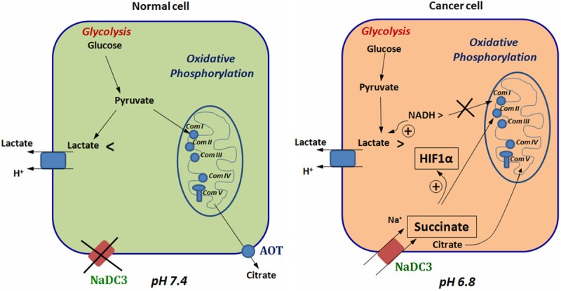
Prostate cancer cells reprogram their metabolism, so that they support their elevated oxidative phosphorylation and promote a cancer friendly microenvironment. This work aimed to explore the mechanisms that cancer cells employ for fueling themselves with energy rich metabolites available in interstitial fluids. The mitochondria oxidative phosphorylation in metastatic prostate cancer DU145 cells and normal prostate epithelial PrEC cells were studied by high-resolution respirometry. An important finding was that prostate cancer cells at acidic pH 6.8 are capable of consuming exogenous succinate, while physiological pH 7.4 was not favorable for this process. Using specific inhibitors, it was demonstrated that succinate is transported in cancer cells by the mechanism of plasma membrane Na(+)-dependent dycarboxylic acid transporter NaDC3 (SLC13A3 gene). Although the level of expression of SLC13A3 was not significantly altered when maintaining cells in the medium with lower pH, the respirometric activity of cells under acidic condition was elevated in the presence of succinate. In contrast, normal prostate cells while expressing NaDC3 mRNA do not produce NaDC3 protein. The mechanism of succinate influx via NaDC3 in metastatic prostate cancer cells could yield a novel target for anti-cancer therapy and has the potential to be used for imaging-based diagnostics to detect non-glycolytic tumors.

Keywords: Na+-dicarboxylate transporter; Prostate cancer; acidic tumor microenvironment; mitochondria oxidative phosphorylation; succinate.


Figures

Figure 1
Figure 1
Succinate uptake by DU145 prostate cancer and PrEC prostate normal cells at different pH. A, B. Representative original records of DU145 cells oxygen consumption measured in the buffer with pH 7.4 and 6.8. After short period of stabilization (a basal respiration, V0) the respiratory enzymes were activated by 40 nM FCCP (VFCCP), and then cells were challenged by 10 mM succinate (VSuc). At the end of the experiment, 10 µM of digitonin was added to provoke a non-transporter-mediated influx of succinate (VDig). C. Oxygen consumption by PrEC cells in the buffer with pH 6.8. D, E. Quantitative data of oxygen consumption rates of DU145 and PrEC cells presented as mean ± S.E.M. (n = 4-6), *p < 0.0468. In DU145 cells digitonin accelerated succinate oxidation is about 12 times higher than in PrEC cells (digit inserts).
Figure 2
Figure 2
Substrate titration analysis of complex II-dependent respiration of DU145 cells. A, B. The representative original oxygraphic records of DU145 cells upon titration with gradually increasing doses of succinate (Suc) are presented. Succinate transport is favored only at acidic condition. The incubation conditions are the same as in legend to Figure 1. C, D. Similar substrate titration protocol applied after inhibition of complex I-mediated respiration with 1 µg/ml rotenone (Rot). Following 40 nM FCCP activation the cells were stimulated by increasing doses of succinate. Digitonin permeabilization of cells further increased the rates of respiration due to a massive succinate influx. Disturbance of cellular content due to digitonin permeabilization results in respiration decline. E. Kinetics of succinate transport presented as a per cent of maximum digitonin-stimulated respiration versus succinate concentrations. Inset is the transporter activity expressed as a per cent of the highest succinate-mediated respiration rates. Data are presented as mean ± S.E.M. (n = 3).
Figure 3
Figure 3
Evaluation of membrane stability of DU145 cells mitochondria upon treatment with FCCP and succinate oxidation by modulation of extracellular calcium content. A. DU145 cells were preloaded with mitochondria membrane potential sensitive dye MitoRed. The background MitoRed intensity is shown as black bars. Cells were exposed to 20 nM (dark grey bars) and 40 nM (light grey bars) FCCP to stimulate their oxidative activities, which restored to its initial level after 15-20 minutes. As a positive control cells were treated with 2 µM FCCP, the dose which eliminates mitochondria membrane potential (white bars). B. DU145 cells succinate oxidation activities measured in the buffers with pH 6.8 containing different concentrations of calcium. The protocol of cell pre-activation with 40 nM FCCP has been applied. The flux control ratios were normalized for FCCP-stimulated respiration. Data are presented as mean ± S.E.M. (n = 4-8). The difference in succinate oxidation rates in the presence of 0.0001 and 0.1 or 0.5 mM CaCl2 was shown to be non-significant. *p < 0.0114, *** p < 0.0008.
Figure 4
Figure 4
Effects of inhibitors on succinate uptake by DU145 prostate cancer cells in acidic buffer. The protocol of FCCP pretreatment was applied as on Figure 1. After short period of stabilization the respiratory enzymes were activated by 40 nM FCCP. A. After mild stimulation, cells were activated by 10 mM succinate. To determine the optimal concentration of the inhibitor, which however preserves the mitochondria intactness, the cells were titrated with gradually increasing doses of mersalyl (10, 20, 60, 120, 180, 240, 300, and 360 µM). Subsequently, 10 µM of digitonin was added to provoke a massive non-transporter-mediated influx of succinate. B. The chosen 250 µM dose of mersalyl was applied before addition of succinate, than cells were permeabilized with digitonin. C. The quantitative data of changes in oxygen consumption rates in the presence of inhibitors are presented as mean ± S.E.M. (n = 4-6). Alternative to 250 µM mersalyl (Mer) dicarboxylic acid transporter inhibitor N-ethylmaleimide (NEM) was used in concentration of 120 µM.
Figure 5
Figure 5
Analysis of dicarboxylic acid transporters transcripts and protein expression profiles in prostate cancer and normal cells. A. RT-PCR and Western blot tests of NaDC3 transporter. Images of polyacrylamide gel containing cDNA reverse-transcribes from DU145 and PrEC cell RNAs along with RNAs prepared from positive control HepG2 and HEK293T/17 cells demonstrate the presence of NaDC3 mRNA in both prostate normal and cancer cells. However, the transporter protein was expressed only in DU145 prostate cancer cells being by 29% down regulated in cells grown in acidic medium. Densitometry values of DC3 protein bands were normalized to a loading venculin control. To calculate a relative ratio the NaDC3 expression level at pH 7.4 was designated as 1, *p < 0.033. B. RT-PCR and Western blot tests of NaCT transporter. Our primers enabled detection of NaCT mRNA expression in both prostate normal and cancer cells. Western blot analysis revealed no expression of the known Na+-dependent citrate transporter in either of investigated prostate cell lines.
Figure 6
Figure 6
Succinate oxidation by different cell lines at pH 6.8 and 7.4. Incubation conditions as in the legend to Figure 1. Respiratory enzymes were activated with 40 nM FCCP and then stimulated with 10 mM succinate. The values of a per cent of FCCP-stimulated oxygen consumption rates presented as mean ± S.E.M. (n = 4-8). *p < 0.038, ** p < 0.006.
Figure 7
Figure 7
The schematics of distinct cross-talk mechanisms between two major energy generating pathways in prostate normal versus cancer cells. Abbreviations: Com I, II, II, IV, V are respiratory system enzymes and ATP synthase, AOT-plasma membrane organic anion transporter, Na+DC3-plasma membrane sodium-dependent dicarboxylic acid transporter, HIF1α-hypoxia inducing factor 1α.

Similar articles

Cited by

References

    1. Swartz MA, Iida N, Roberts EW, Sangaletti S, Wong MH, Yull FE, Coussens LM, DeClerck YA. Tumor microenvironment complexity: emerging roles in cancer therapy. Cancer Res. 2012;72:2473–2480. - PMC - PubMed
    1. Martinez-Outschoorn UE, Pestell RG, Howell A, Tykocinski ML, Nagajyothi F, Machado FS, Tanowitz HB, Sotgia F, Lisanti MP. Energy transfer in “parasitic” cancer metabolism: mitochondria are the powerhouse and Achilles’ heel of tumor cells. Cell Cycle. 2011;10:4208–4216. - PMC - PubMed
    1. Edlund M, Sung SY, Chung LW. Modulation of prostate cancer growth in bone microenvironments. J Cell Biochem. 2004;91:686–705. - PubMed
    1. Robey IF, Baggett BK, Kirkpatrick ND, Roe DJ, Dosescu J, Sloane BF, Hashim AI, Morse DL, Raghunand N, Gatenby RA, Gillies RJ. Bicarbonate increases tumor pH and inhibits spontaneous metastases. Cancer Res. 2009;69:2260–2268. - PMC - PubMed
    1. Kato Y, Ozawa S, Miyamoto C, Maehata Y, Suzuki A, Maeda T, Baba Y. Acidic extracellular microenvironment and cancer. Cancer Cell Int. 2013;13:89. - PMC - PubMed

LinkOut - more resources