

Proinflammatory Mediators Enhance the Osteogenesis of Human Mesenchymal Stem Cells after Lineage Commitment
- PMID: 26176237
- PMCID: PMC4503569
- DOI: 10.1371/journal.pone.0132781
Proinflammatory Mediators Enhance the Osteogenesis of Human Mesenchymal Stem Cells after Lineage Commitment
Abstract
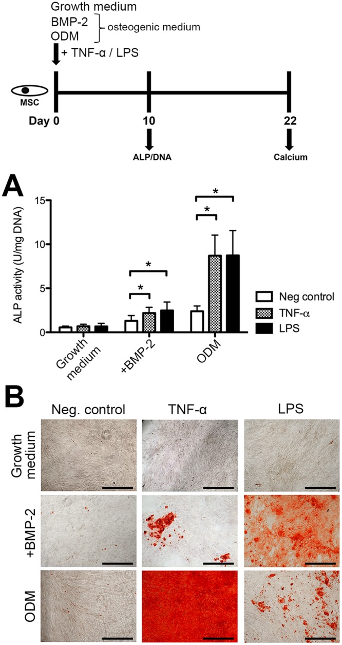
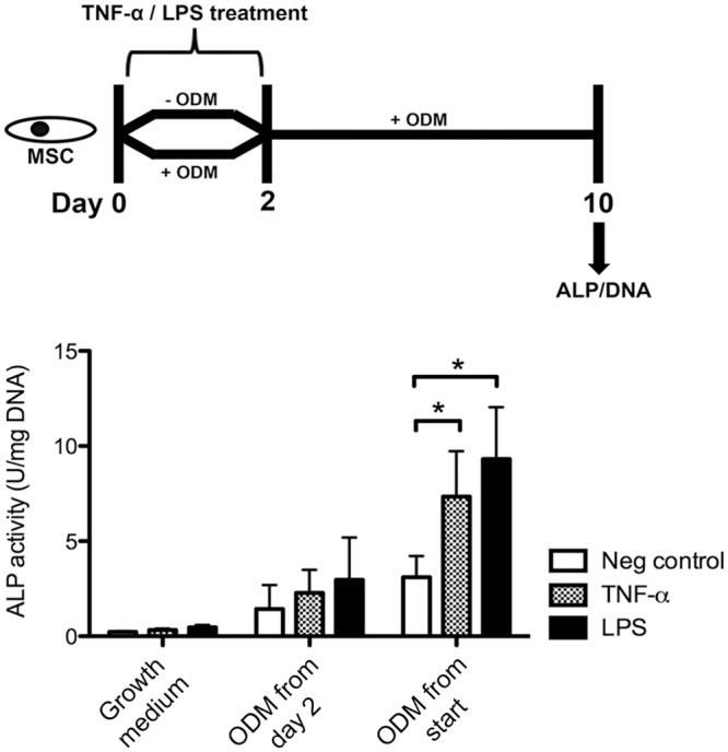
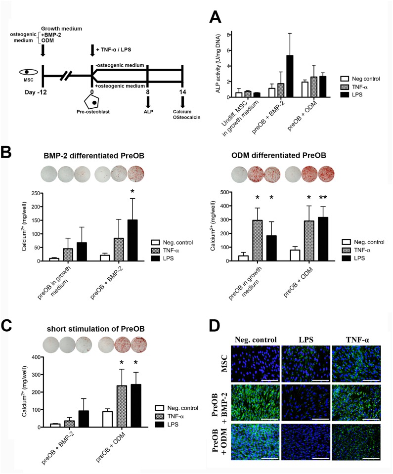
Several inflammatory processes underlie excessive bone formation, including chronic inflammation of the spine, acute infections, or periarticular ossifications after trauma. This suggests that local factors in these conditions have osteogenic properties. Mesenchymal stem cells (MSCs) and their differentiated progeny contribute to bone healing by synthesizing extracellular matrix and inducing mineralization. Due to the variation in experimental designs used in vitro, there is controversy about the osteogenic potential of proinflammatory factors on MSCs. Our goal was to determine the specific conditions allowing the pro-osteogenic effects of distinct inflammatory stimuli. Human bone marrow MSCs were exposed to tumor necrosis factor alpha (TNF-α) and lipopolysaccharide (LPS). Cells were cultured in growth medium or osteogenic differentiation medium. Alternatively, bone morphogenetic protein 2 (BMP-2) was used as osteogenic supplement to simulate the conditions in vivo. Alkaline phosphatase activity and calcium deposition were indicators of osteogenicity. To elucidate lineage commitment-dependent effects, MSCs were pre-differentiated prior treatment. Our results show that TNF-α and LPS do not affect the expression of osteogenic markers by MSCs in the absence of an osteogenic supplement. In osteogenic differentiation medium or together with BMP-2 however, these mediators highly stimulated their alkaline phosphatase activity and subsequent matrix mineralization. In pre-osteoblasts, matrix mineralization was significantly increased by these mediators, but irrespective of the culture conditions. Our study shows that inflammatory factors potently enhance the osteogenic capacity of MSCs. These properties may be harnessed in bone regenerative strategies. Importantly, the commitment of MSCs to the osteogenic lineage greatly enhances their responsiveness to inflammatory signals.
Conflict of interest statement
Figures
Similar articles
-
CD200 expression in human cultured bone marrow mesenchymal stem cells is induced by pro-osteogenic and pro-inflammatory cues.J Cell Mol Med. 2016 Apr;20(4):655-65. doi: 10.1111/jcmm.12752. Epub 2016 Jan 16. J Cell Mol Med. 2016. PMID: 26773707 Free PMC article.
-
Bortezomib enhances the osteogenic differentiation capacity of human mesenchymal stromal cells derived from bone marrow and placental tissues.Biochem Biophys Res Commun. 2014 May 16;447(4):580-5. doi: 10.1016/j.bbrc.2014.04.044. Epub 2014 Apr 18. Biochem Biophys Res Commun. 2014. PMID: 24747566
-
Paracrine effect of inflammatory cytokine-activated bone marrow mesenchymal stem cells and its role in osteoblast function.J Biosci Bioeng. 2016 Feb;121(2):213-9. doi: 10.1016/j.jbiosc.2015.05.017. Epub 2015 Aug 25. J Biosci Bioeng. 2016. PMID: 26315505
-
[Advances in osteogenic mechanism and osteogenic effects of bone morphogenetic protein 6].Zhongguo Xiu Fu Chong Jian Wai Ke Za Zhi. 2013 Sep;27(9):1144-7. Zhongguo Xiu Fu Chong Jian Wai Ke Za Zhi. 2013. PMID: 24279032 Review. Chinese.
-
The Influence of Inflammatory Cytokines on the Proliferation and Osteoblastic Differentiation of MSCs.Curr Stem Cell Res Ther. 2017;12(5):401-408. doi: 10.2174/1574888X12666170509102222. Curr Stem Cell Res Ther. 2017. PMID: 28486918 Review.
Cited by
-
NF-κB decoy oligodeoxynucleotide mitigates wear particle-associated bone loss in the murine continuous infusion model.Acta Biomater. 2016 Sep 1;41:273-81. doi: 10.1016/j.actbio.2016.05.038. Epub 2016 May 31. Acta Biomater. 2016. PMID: 27260104 Free PMC article.
-
Aesculetin Accelerates Osteoblast Differentiation and Matrix-Vesicle-Mediated Mineralization.Int J Mol Sci. 2021 Nov 17;22(22):12391. doi: 10.3390/ijms222212391. Int J Mol Sci. 2021. PMID: 34830274 Free PMC article.
-
Hyperbaric Oxygen Therapy Improves the Osteogenic and Vasculogenic Properties of Mesenchymal Stem Cells in the Presence of Inflammation In Vitro.Int J Mol Sci. 2020 Feb 20;21(4):1452. doi: 10.3390/ijms21041452. Int J Mol Sci. 2020. PMID: 32093391 Free PMC article.
-
The crosstalk between vascular MSCs and inflammatory mediators determines the pro-calcific remodelling of human atherosclerotic aneurysm.Stem Cell Res Ther. 2017 Apr 26;8(1):99. doi: 10.1186/s13287-017-0554-x. Stem Cell Res Ther. 2017. PMID: 28446225 Free PMC article.
-
Bioengineering Approaches to Accelerate Clinical Translation of Stem Cell Therapies Treating Osteochondral Diseases.Stem Cells Int. 2020 Dec 24;2020:8874742. doi: 10.1155/2020/8874742. eCollection 2020. Stem Cells Int. 2020. PMID: 33424981 Free PMC article. Review.
References
-
- Arrington ED, Smith WJ, Chambers HG, Bucknell AL, Davino NA. Complications of iliac crest bone graft harvesting. Clinical orthopaedics and related research. 1996: 300–309. - PubMed
-
- LeGeros RZ. Properties of osteoconductive biomaterials: calcium phosphates. Clinical orthopaedics and related research. 2002: 81–98. - PubMed
-
- Petite H, Viateau V, Bensaid W, Meunier A, de Pollak C, Bourguignon M, et al. Tissue-engineered bone regeneration. Nature biotechnology. 2000; 18: 959–963. - PubMed
-
- Carragee EJ, Hurwitz EL, Weiner BK. A critical review of recombinant human bone morphogenetic protein-2 trials in spinal surgery: emerging safety concerns and lessons learned. The spine journal: official journal of the North American Spine Society. 2011; 11: 471–491. - PubMed
-
- Geiger M, Li RH, Friess W. Collagen sponges for bone regeneration with rhBMP-2. Advanced drug delivery reviews. 2003; 55: 1613–1629. - PubMed
MeSH terms
Substances
LinkOut - more resources
Full Text Sources
Other Literature Sources
