

Histidine-rich calcium binding protein promotes growth of hepatocellular carcinoma in vitro and in vivo
- PMID: 26176291
- PMCID: PMC4638025
- DOI: 10.1111/cas.12743
Histidine-rich calcium binding protein promotes growth of hepatocellular carcinoma in vitro and in vivo
Abstract
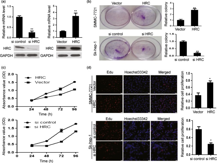
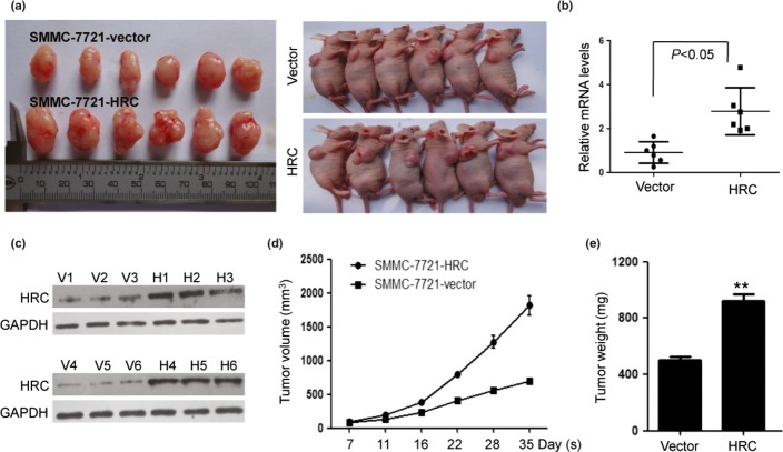
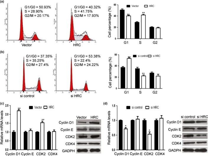
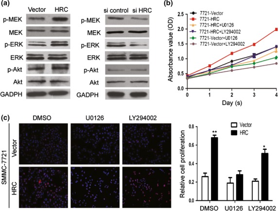
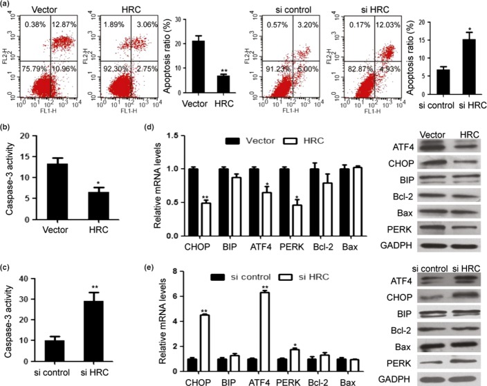
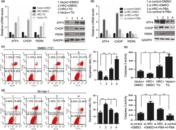
We have recently shown that the histidine-rich calcium binding protein (HRC) promotes the invasion and metastasis of hepatocellular carcinoma (HCC). In the current study, we evaluated whether HRC may also affect the growth of HCC. We found that ectopic expression of HRC obviously enhanced proliferation and colony formation, while suppression of HRC exhibited inhibitory effects. Furthermore, we demonstrated that HRC promoted tumor growth in nude mice. These effects may result from the ability of HRC to upregulate cyclinD1 and cyclin-dependent kinase 2 (CDK2) expressions and promote G1/S transition. Further study showed that MEK/ERK signaling pathway was involved in HRC-induced cell proliferation. Interestingly, overexpression or depletion of HRC revealed its regulation on endoplasmic reticulum stress (ERS) and apoptosis, which was partially dependent on PERK/ATF4/CHOP signaling pathway. In addition, blocking ERS using 4-phenylbutyric acid (4-PBA) not only downregulated the expression of PERK, ATF4 and CHOP, but also significantly decreased apoptosis induced by HRC silence, whereas ERS inducer thapsigargin (TG) exerted the opposite effects. Our study thus demonstrates a role of HRC in promoting HCC growth, besides its role in inducing HCC metastasis, and highlights HRC as a promising intervention target for HCC.
Keywords: Apoptosis; endoplasmic reticulum stress; hepatocellular carcinoma; histidine-rich calcium binding protein; proliferation.
© 2015 The Authors. Cancer Science published by Wiley Publishing Asia Pty Ltd on behalf of Japanese Cancer Association.
Figures
Similar articles
-
HRC promotes anoikis resistance and metastasis by suppressing endoplasmic reticulum stress in hepatocellular carcinoma.Int J Med Sci. 2021 Jun 26;18(14):3112-3124. doi: 10.7150/ijms.60610. eCollection 2021. Int J Med Sci. 2021. PMID: 34400882 Free PMC article.
-
The histidine-rich calcium binding protein (HRC) promotes tumor metastasis in hepatocellular carcinoma and is upregulated by SATB1.Oncotarget. 2015 Mar 30;6(9):6811-24. doi: 10.18632/oncotarget.3049. Oncotarget. 2015. PMID: 25762622 Free PMC article.
-
β-Elemonic acid inhibits the growth of human Osteosarcoma through endoplasmic reticulum (ER) stress-mediated PERK/eIF2α/ATF4/CHOP activation and Wnt/β-catenin signal suppression.Phytomedicine. 2020 Apr;69:153183. doi: 10.1016/j.phymed.2020.153183. Epub 2020 Feb 7. Phytomedicine. 2020. PMID: 32113150
-
ATP citrate lyase inhibitor triggers endoplasmic reticulum stress to induce hepatocellular carcinoma cell apoptosis via p-eIF2α/ATF4/CHOP axis.J Cell Mol Med. 2021 Feb;25(3):1468-1479. doi: 10.1111/jcmm.16235. Epub 2021 Jan 3. J Cell Mol Med. 2021. PMID: 33393219 Free PMC article.
-
Cartilage-Specific Autophagy Deficiency Promotes ER Stress and Impairs Chondrogenesis in PERK-ATF4-CHOP-Dependent Manner.J Bone Miner Res. 2017 Oct;32(10):2128-2141. doi: 10.1002/jbmr.3134. Epub 2017 Apr 14. J Bone Miner Res. 2017. PMID: 28304100
Cited by
-
The combination of hydroxychloroquine and 2-deoxyglucose enhances apoptosis in breast cancer cells by blocking protective autophagy and sustaining endoplasmic reticulum stress.Cell Death Discov. 2022 Jun 11;8(1):286. doi: 10.1038/s41420-022-01074-6. Cell Death Discov. 2022. PMID: 35690609 Free PMC article.
-
GC-MS-based untargeted metabolic profiling of malignant mesothelioma plasma.PeerJ. 2023 May 18;11:e15302. doi: 10.7717/peerj.15302. eCollection 2023. PeerJ. 2023. PMID: 37220527 Free PMC article.
-
Inhibition of lung cancer by vitamin D depends on downregulation of histidine-rich calcium-binding protein.J Adv Res. 2020 Aug 27;29:13-22. doi: 10.1016/j.jare.2020.08.013. eCollection 2021 Mar. J Adv Res. 2020. PMID: 33842001 Free PMC article.
-
Apoptotic-like PCD inducing HRC gene when silenced enhances multiple disease resistance in plants.Sci Rep. 2022 Nov 27;12(1):20402. doi: 10.1038/s41598-022-24831-0. Sci Rep. 2022. PMID: 36437285 Free PMC article.
-
Knockdown of KIAA1199 attenuates growth and metastasis of hepatocellular carcinoma.Cell Death Discov. 2018 Nov 12;4:102. doi: 10.1038/s41420-018-0099-5. eCollection 2018. Cell Death Discov. 2018. PMID: 30455988 Free PMC article.
References
Publication types
MeSH terms
Substances
LinkOut - more resources
Full Text Sources
Other Literature Sources
Medical
Research Materials
Miscellaneous
