

Osteopontin is proteolytically processed by matrix metalloproteinase 9
- PMID: 26176332
- PMCID: PMC4764993
- DOI: 10.1139/cjpp-2015-0019
Osteopontin is proteolytically processed by matrix metalloproteinase 9
Abstract
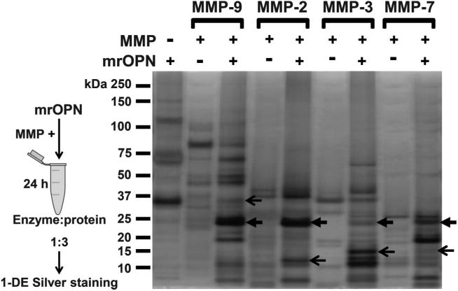
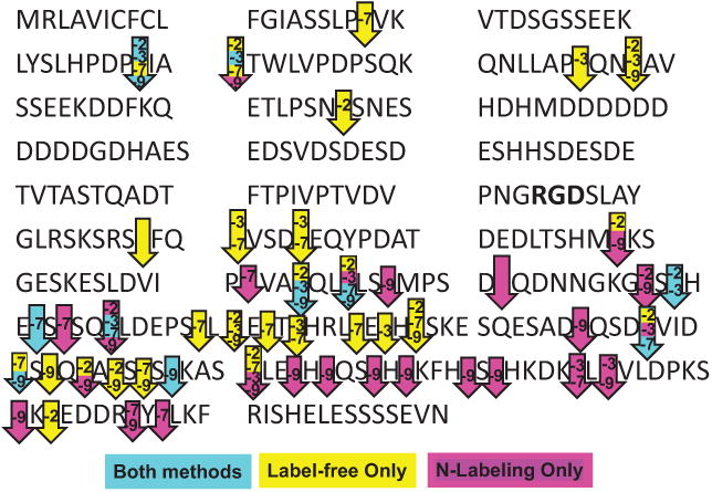
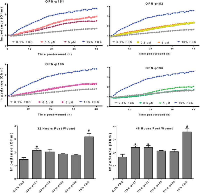
Osteopontin is robustly upregulated following myocardial infarction (MI), which suggests that it has an important role in post-MI remodeling of the left ventricle (LV). Osteopontin deletion results in increased LV dilation and worsened cardiac function. Thus, osteopontin exerts protective effects post-MI, but the mechanisms have yet to be defined. Matrix metalloproteinases (MMPs) regulate LV remodeling post-MI, and osteopontin is a known substrate for MMP-2, -3, -7, and -9, although the cleavage sites have not been mapped. Osteopontin-derived peptides can exert distinct biological functions that may depend on their cleavage sites. We mapped the MMP-9 cleavage sites via LC-MS/MS analysis using label-free and N-terminal labeling methods, and compared them with those of MMP-2, -3, and -7. Each MMP yielded a unique cleavage profile with few overlapping cleavage sites. Using synthetic peptides, we validated 3 sites for MMP-9 cleavage at amino acid positions 151-152, 193-194, and 195-196. Four peptides were synthesized based on the upstream- and downstream-generated fragments and were tested for biological activity in isolated cardiac fibroblasts. Two peptides increased cardiac fibroblast migration rates post-wounding (p < 0.05 compared with the negative control). Our study highlights the importance of osteopontin processing, and confirms that different cleavage sites generate osteopontin peptides with distinct biological functions.
Keywords: MMP-9; MPM-9; cardiac; cardiaque; cleavage sites; fibroblastes; fibroblasts; infarctus du myocarde; mass spectrometry; matrix metalloproteinases; myocardial infarction; métalloprotéinases matricielles; osteopontin; ostéopontine; peptides; proteomics; protéomique; sites de clivage; spectométrie de masse.
Figures


Similar articles
-
A Novel Collagen Matricryptin Reduces Left Ventricular Dilation Post-Myocardial Infarction by Promoting Scar Formation and Angiogenesis.J Am Coll Cardiol. 2015 Sep 22;66(12):1364-74. doi: 10.1016/j.jacc.2015.07.035. J Am Coll Cardiol. 2015. PMID: 26383724 Free PMC article.
-
Inhibition of matrix metalloproteinases improves left ventricular function in mice lacking osteopontin after myocardial infarction.Mol Cell Biochem. 2009 Feb;322(1-2):53-62. doi: 10.1007/s11010-008-9939-6. Epub 2008 Nov 1. Mol Cell Biochem. 2009. PMID: 18979185 Free PMC article.
-
Plasma MMP-2, MMP-9 and N-BNP in long-term survivors following complicated myocardial infarction: relation to cardiac magnetic resonance imaging measures of left ventricular structure and function.J Card Fail. 2007 Dec;13(10):843-9. doi: 10.1016/j.cardfail.2007.07.006. J Card Fail. 2007. PMID: 18068618 Clinical Trial.
-
The circular relationship between matrix metalloproteinase-9 and inflammation following myocardial infarction.IUBMB Life. 2015 Aug;67(8):611-8. doi: 10.1002/iub.1408. Epub 2015 Aug 13. IUBMB Life. 2015. PMID: 26269290 Free PMC article. Review.
-
MMP-9 signaling in the left ventricle following myocardial infarction.Am J Physiol Heart Circ Physiol. 2016 Jul 1;311(1):H190-8. doi: 10.1152/ajpheart.00243.2016. Epub 2016 May 20. Am J Physiol Heart Circ Physiol. 2016. PMID: 27208160 Free PMC article. Review.
Cited by
-
Extracellular matrix roles in cardiorenal fibrosis: Potential therapeutic targets for CVD and CKD in the elderly.Pharmacol Ther. 2019 Jan;193:99-120. doi: 10.1016/j.pharmthera.2018.08.014. Epub 2018 Aug 25. Pharmacol Ther. 2019. PMID: 30149103 Free PMC article. Review.
-
Interactions and Trends of Interleukins, PAI-1, CRP, and TNF-α in Inflammatory Responses during the Perioperative Period of Joint Arthroplasty: Implications for Pain Management-A Narrative Review.J Pers Med. 2024 May 17;14(5):537. doi: 10.3390/jpm14050537. J Pers Med. 2024. PMID: 38793119 Free PMC article. Review.
-
MMP-3-mediated cleavage of OPN is involved in copper oxide nanoparticle-induced activation of fibroblasts.Part Fibre Toxicol. 2023 May 22;20(1):22. doi: 10.1186/s12989-023-00532-y. Part Fibre Toxicol. 2023. PMID: 37217992 Free PMC article.
-
The Multifaceted Role of Osteopontin in Prostate Pathologies.Biomedicines. 2023 Oct 26;11(11):2895. doi: 10.3390/biomedicines11112895. Biomedicines. 2023. PMID: 38001899 Free PMC article. Review.
-
Assigning matrix metalloproteinase roles in ischaemic cardiac remodelling.Nat Rev Cardiol. 2018 Aug;15(8):471-479. doi: 10.1038/s41569-018-0022-z. Nat Rev Cardiol. 2018. PMID: 29752454 Free PMC article. Review.
References
Publication types
MeSH terms
Substances
Grants and funding
LinkOut - more resources
Full Text Sources
Other Literature Sources
Research Materials
Miscellaneous
