. 2015 Aug;19(8):1783-94.
doi: 10.1111/jcmm.12624. Epub 2015 Jul 14.

The secretome of myocardial telocytes modulates the activity of cardiac stem cells

Affiliations

The secretome of myocardial telocytes modulates the activity of cardiac stem cells

Radu Albulescu et al. J Cell Mol Med. 2015 Aug.

Abstract

Telocytes (TCs) are interstitial cells that are present in numerous organs, including the heart interstitial space and cardiac stem cell niche. TCs are completely different from fibroblasts. TCs release extracellular vesicles that may interact with cardiac stem cells (CSCs) via paracrine effects. Data on the secretory profile of TCs and the bidirectional shuttle vesicular signalling mechanism between TCs and CSCs are scarce. We aimed to characterize and understand the in vitro effect of the TC secretome on CSC fate. Therefore, we studied the protein secretory profile using supernatants from mouse cultured cardiac TCs. We also performed a comparative secretome analysis using supernatants from rat cultured cardiac TCs, a pure CSC line and TCs-CSCs in co-culture using (i) high-sensitivity on-chip electrophoresis, (ii) surface-enhanced laser desorption/ionization time-of-flight mass spectrometry and (iii) multiplex analysis by Luminex-xMAP. We identified several highly expressed molecules in the mouse cardiac TC secretory profile: interleukin (IL)-6, VEGF, macrophage inflammatory protein 1α (MIP-1α), MIP-2 and MCP-1, which are also present in the proteome of rat cardiac TCs. In addition, rat cardiac TCs secrete a slightly greater number of cytokines, IL-2, IL-10, IL-13 and some chemokines like, GRO-KC. We found that VEGF, IL-6 and some chemokines (all stimulated by IL-6 signalling) are secreted by cardiac TCs and overexpressed in co-cultures with CSCs. The expression levels of MIP-2 and MIP-1α increased twofold and fourfold, respectively, when TCs were co-cultured with CSCs, while the expression of IL-2 did not significantly differ between TCs and CSCs in mono culture and significantly decreased (twofold) in the co-culture system. These data suggest that the TC secretome plays a modulatory role in stem cell proliferation and differentiation.

Keywords: Luminex-xMAP; SELDI-TOF; cardiac stem cells; chemokines; cytokines; fibroblasts; growth factors; telocytes.


Figures

Figure 1
Figure 1
Protein separation of cell-free supernatants from cultures (three subsequent passages) of mouse TCs by on-chip electrophoresis. The bands at 10.3, 13.2, 22.5, 119.2 and 129.5 kD were present in all mouse TCs passages, but not in FB culture. TC-P1 – first passage 1, TC-P2 – second passage, TC-P3 – third passage, FB – fibroblasts, Lad – ladder that illustrates the molecular weight markers.
Figure 2
Figure 2
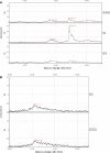
Protein profiling analysis of TC and FB supernatants by surface-enhanced laser desorption and ionization mass spectrometry. Characteristic proteins that are secreted by TCs were identified at m/z values of ∼11.7 kD, from 13.8 to 14.8, and at 22.5 kD. The peaks suggest the presence of MIP-1α, MIP-2 dimer, MCP-1 and VEGF. m/z range: 0–25,000 D, CM10 chips, SPA matrix. Control – cell culture medium, TC-P1 – first passage, TC-P2 – second passage, TC-P3 – third passage, FB – fibroblasts.
Figure 3
Figure 3
The levels of IL 6 cytokine in the serum-free culture supernatants of mouse myocardial TCs compared to 3T3 fibroblasts as determined by xMAP technology. The data are expressed as the mean ± SE from three independent experiments and were analysed with Student’s t-test. TC1 – first passage mouse TCs, TC2 – second passage mouse TCs, TC3 – third passage mouse TCs, FB: fibroblasts.
Figure 4
Figure 4
VEGF expression levels in the cell culture supernatants of mouse myocardial TCs compared with 3T3 fibroblasts. (A) VEGF levels in serum-free supernatants. (B) VEGF levels in serum-supplemented media. TC1 – first passage TCs, TC2 – second passage TCs, TC3 – third passage TCs, FB – fibroblasts.
Figure 5
Figure 5
(A) Proteins that were secreted in the cell culture supernatants of rat myocardial TCs as assessed by SELDI-TOF-MS analysis. A significant enhancement of the 14.8 kD peak in TC mono-culture compared with CSC mono-culture and TC-CSC co-culture is evident. m/z range: 10,000–20,000 D, CM10 chips, SPA matrix. (B) Outline of the 22.2 kD peak (IL-6) in the SELDI-TOF-MS spectra of proteins that were secreted by rat TCs. CM10 chips, SPA matrix. The expression of the protein associated with this peak does not significantly differ between the TC and TC-CSC co-cultures.
Figure 6
Figure 6
Luminex-xMAP detection of secreted cyto- and chemokines in the cell culture supernatants of rat myocardial TC mono-, CSC mono- and TC-CSC co-cultures: (A) IL-6, (B) MIP-2, (C) MIP-1α, (D) MCP-1. The results were normalized to cell counts. P < 0.05, n = 3.

Similar articles

Cited by

References

    1. Popescu LM, Faussone-Pellegrini MS. TELOCYTES - a case of serendipity: the winding way from Interstitial Cells of Cajal (ICC), via Interstitial Cajal-Like Cells (ICLC) to TELOCYTES. J Cell Mol Med. 2010;14:729–40. - PMC - PubMed
    1. Suciu L, Nicolescu MI, Popescu LM. Cardiac telocytes: serial dynamic images in cell culture. J Cell Mol Med. 2010;14:2687–92. - PMC - PubMed
    1. Gherghiceanu M, Popescu LM. Cardiac telocytes - their junctions and functional implications. Cell Tissue Res. 2012;348:265–79. - PMC - PubMed
    1. Cretoiu D, Hummel E, Zimmermann H, et al. Human cardiac telocytes: 3D imaging by FIB-SEM tomography. J Cell Mol Med. 2014;18:2157–64. - PMC - PubMed
    1. Yang Y, Sun W, Wu SM, et al. Telocytes in human heart valves. J Cell Mol Med. 2014;18:759–65. - PMC - PubMed

Publication types