

Quantitative LC-MS/MS Analysis of Proteins Involved in Metastasis of Breast Cancer
- PMID: 26176947
- PMCID: PMC4503764
- DOI: 10.1371/journal.pone.0130760
Quantitative LC-MS/MS Analysis of Proteins Involved in Metastasis of Breast Cancer
Abstract
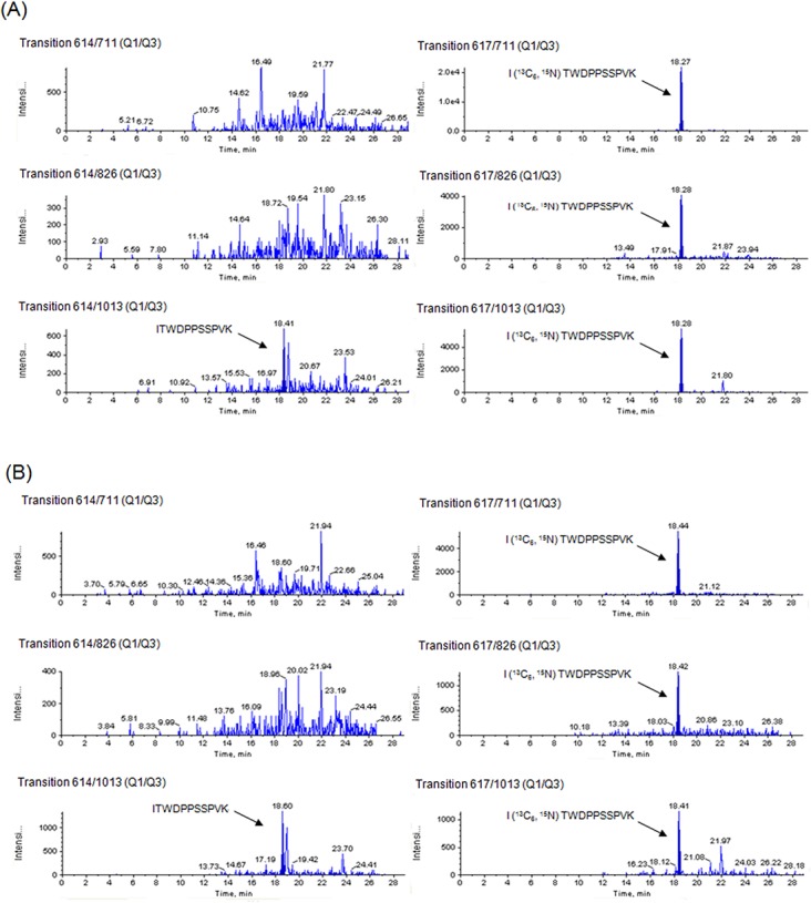
The purpose of this study was to develop quantitative liquid chromatography-tandem mass spectrometry (LC-MS/MS) methods for the analysis of proteins involved in metastasis of breast cancer for diagnosis and determining disease prognosis, as well as to further our understand of metastatic mechanisms. We have previously demonstrated that the protein type XIV collagen may be specifically expressed in metastatic tissues by two dimensional LC-MS/MS. In this study, we developed quantitative LC-MS/MS methods for type XIV collagen. Type XIV collagen was quantified by analyzing 2 peptides generated by digesting type XIV collagen using stable isotope-labeled peptides. The individual concentrations were equivalent between 2 different peptides of type XIV collagen by evaluation of imprecise transitions and using the best transition for the peptide concentration. The results indicated that type XIV collagen is highly expressed in metastatic tissues of patients with massive lymph node involvement compared to non-metastatic tissues. These findings were validated by quantitative real-time RT-PCR. Further studies on type XIV collagen are desired to verify its role as a prognostic factor and diagnosis marker for metastasis.
Conflict of interest statement
Figures


Similar articles
-
Biomarker discovery in low-grade breast cancer using isobaric stable isotope tags and two-dimensional liquid chromatography-tandem mass spectrometry (iTRAQ-2DLC-MS/MS) based quantitative proteomic analysis.J Proteome Res. 2009 Jan;8(1):362-73. doi: 10.1021/pr800622b. J Proteome Res. 2009. PMID: 19053527
-
Internal standards in the quantitative determination of protein biopharmaceuticals using liquid chromatography coupled to mass spectrometry.J Chromatogr B Analyt Technol Biomed Life Sci. 2012 Apr 15;893-894:1-14. doi: 10.1016/j.jchromb.2012.02.021. Epub 2012 Feb 21. J Chromatogr B Analyt Technol Biomed Life Sci. 2012. PMID: 22426285 Review.
-
Comparison of liquid chromatography-tandem mass spectrometry-based targeted proteomics and conventional analytical methods for the determination of P-glycoprotein in human breast cancer cells.J Chromatogr B Analyt Technol Biomed Life Sci. 2013 Oct 1;936:18-24. doi: 10.1016/j.jchromb.2013.07.023. Epub 2013 Aug 3. J Chromatogr B Analyt Technol Biomed Life Sci. 2013. PMID: 23968647
-
A quantitative LC-MS/MS method for comparative analysis of capture-antibody affinity toward protein antigens.J Chromatogr B Analyt Technol Biomed Life Sci. 2011 Sep 15;879(26):2726-32. doi: 10.1016/j.jchromb.2011.07.037. Epub 2011 Aug 2. J Chromatogr B Analyt Technol Biomed Life Sci. 2011. PMID: 21856254
-
Androgen glucuronides analysis by liquid chromatography tandem-mass spectrometry: could it raise new perspectives in the diagnostic field of hormone-dependent malignancies?J Chromatogr B Analyt Technol Biomed Life Sci. 2013 Dec 1;940:24-34. doi: 10.1016/j.jchromb.2013.09.022. Epub 2013 Sep 27. J Chromatogr B Analyt Technol Biomed Life Sci. 2013. PMID: 24140653 Review.
Cited by
-
Identification of Key Genes Associated with Brain Metastasis from Breast Cancer: A Bioinformatics Analysis.Med Sci Monit. 2022 Mar 17;28:e935071. doi: 10.12659/MSM.935071. Med Sci Monit. 2022. PMID: 35296631 Free PMC article.
-
The Role of Tumor Microenvironment in Chemoresistance: 3D Extracellular Matrices as Accomplices.Int J Mol Sci. 2018 Sep 20;19(10):2861. doi: 10.3390/ijms19102861. Int J Mol Sci. 2018. PMID: 30241395 Free PMC article.
-
Quantitative extracellular matrix proteomics to study mammary and liver tissue microenvironments.Int J Biochem Cell Biol. 2016 Dec;81(Pt A):223-232. doi: 10.1016/j.biocel.2016.10.014. Epub 2016 Oct 24. Int J Biochem Cell Biol. 2016. PMID: 27771439 Free PMC article.
-
Primary breast tumor induced extracellular matrix remodeling in premetastatic lungs.Sci Rep. 2023 Oct 30;13(1):18566. doi: 10.1038/s41598-023-45832-7. Sci Rep. 2023. PMID: 37903851 Free PMC article.
-
Premetastatic niche mechanics and organotropism in breast cancer.NPJ Biol Phys Mech. 2025;2(1):11. doi: 10.1038/s44341-025-00015-5. Epub 2025 Apr 3. NPJ Biol Phys Mech. 2025. PMID: 40191104 Free PMC article. Review.
References
-
- Nemoto T, Vana J, Bedwani RN, Baker HW, McGregor FH, Murphy GP. Management and survival of female breast cancer: results of a national survey by the American College of Surgeons. Cancer 1980; 45(12): 2917–2924. - PubMed
-
- SEER Cancer Statistics Review (CSR) 1975–2010. Available: http://seer.cancer.gov/csr/1975_2010/browse_csr.php?sectionSEL=4&pageSEL...
-
- Nakamura Y, Yasuoka H, Tsujimoto M, Yang Q, Imabun S, Nakahara M, et al. Flt-4-positive vessel density correlates with vascular endothelial growth factor-d expression, nodal status, and prognosis in breast cancer. Clin Cancer Res. 2003; 9(14):5313–5317. - PubMed
-
- Goto R, Nakamura Y, Shioyama S, Sanke T, Tozuka Z. Screening of Specific Proteins Involved in Breast Cancer Metastasis by 2D LC-ESI-MS/MS. J. Wakayama Med. Soc. 2014; 65(3)84–89.
MeSH terms
Substances
LinkOut - more resources
Full Text Sources
Other Literature Sources
Medical
