. 2015 Jul 15;10(7):e0133056.
doi: 10.1371/journal.pone.0133056. eCollection 2015.

Versican V1 Overexpression Induces a Myofibroblast-Like Phenotype in Cultured Fibroblasts

Affiliations

Versican V1 Overexpression Induces a Myofibroblast-Like Phenotype in Cultured Fibroblasts

Jon M Carthy et al. PLoS One. .

Abstract

Background: Versican, a chondroitin sulphate proteoglycan, is one of the key components of the provisional extracellular matrix expressed after injury. The current study evaluated the hypothesis that a versican-rich matrix alters the phenotype of cultured fibroblasts.

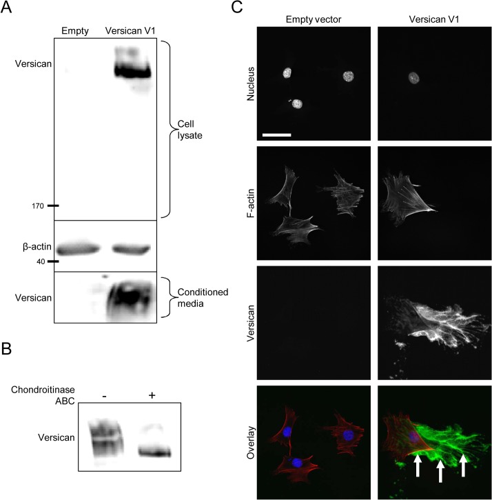
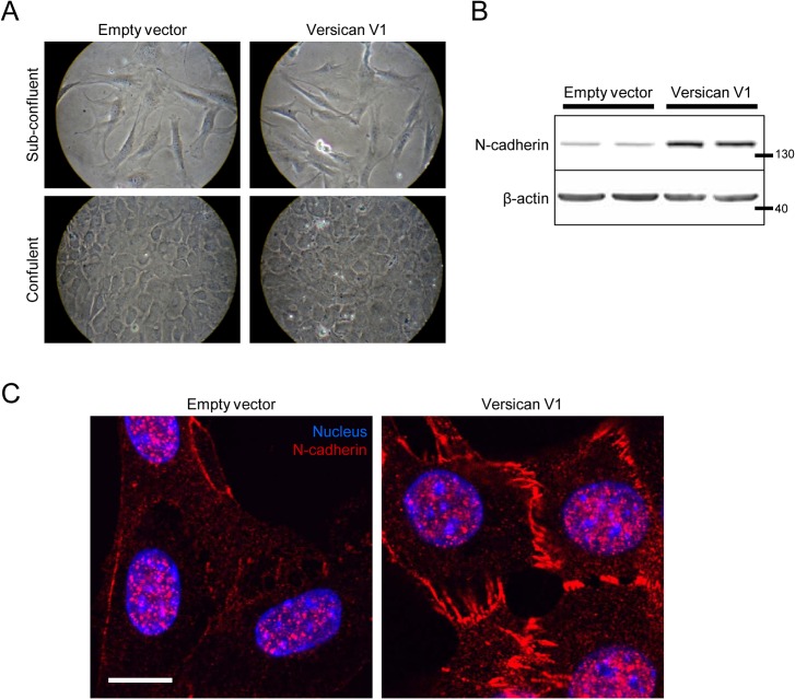
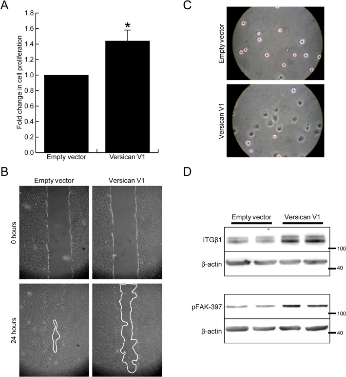
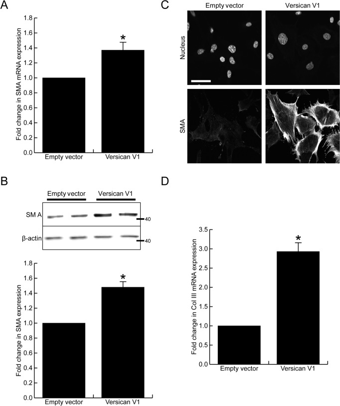
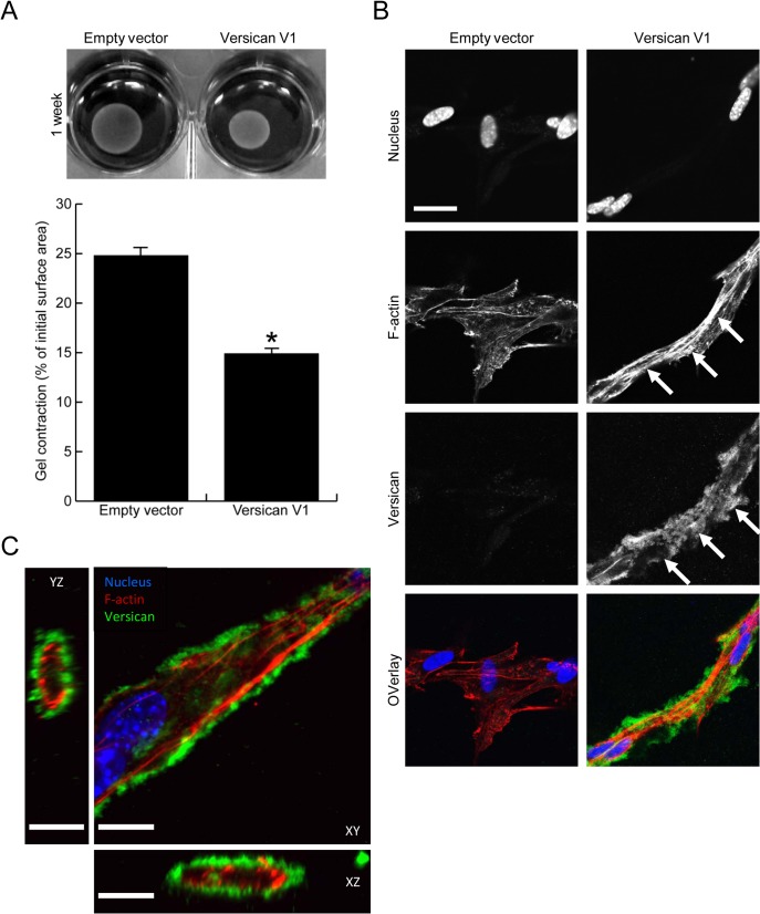
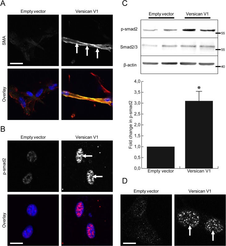
Methods and results: The full-length cDNA for the V1 isoform of human versican was cloned and the recombinant proteoglycan was expressed in murine fibroblasts. Versican expression induced a marked change in fibroblast phenotype. Functionally, the versican-expressing fibroblasts proliferated faster and displayed enhanced cell adhesion, but migrated slower than control cells. These changes in cell function were associated with greater N-cadherin and integrin β1 expression, along with increased FAK phosphorylation. The versican-expressing fibroblasts also displayed expression of smooth muscle α-actin, a marker of myofibroblast differentiation. Consistent with this observation, the versican fibroblasts displayed increased synthetic activity, as measured by collagen III mRNA expression, as well as a greater capacity to contract a collagen lattice. These changes appear to be mediated, at least in part, by an increase in active TGF-β signaling in the versican expressing fibroblasts, and this was measured by phosphorylation and nuclear accumulation of SMAD2.

Conclusions: Collectively, these data indicate versican expression induces a myofibroblast-like phenotype in cultured fibroblasts.


Conflict of interest statement

Competing Interests: The authors have declared that no competing interests exist.

Figures

Fig 1
Fig 1. Versican expression in murine fibroblasts.
(A) Western blot of recombinant versican expression in the cell lysate and conditioned medium of versican-transfected fibroblasts, suggesting the recombinant protein is synthesized and secreted. (B) Cell lysates were digested with chondroitinase ABC prior to Western blotting to confirm the presence of GAG chains on the recombinant versican. In the absence of chondroitinase, versican appeared as a large smear representing molecules of different molecular weights; after chondroitinase treatment, versican appeared as a compact band at the size of the smallest versican molecules from the smear, suggesting the GAG chains were present and had been removed. (C) Immunofluorescence microscopy showed recombinant versican was deposited into the ECM in versican-transfected cells (green, arrows). (Scale bar = 47.00 μm).
Fig 2
Fig 2. Versican increases N-cadherin expression.
(A) Light microscopy shows no major change in cell morphology in versican-transfected fibroblasts at both sub-confluent and confluent densities. (B) Representative Western blot showing increased expression of N-cadherin in versican-transfected cells. (C) Confocal microscopy confirmed the increased N-cadherin expression in versican-transfected cells. (Scale bar = 12.00 μm).
Fig 3
Fig 3. Versican alters the function of fibroblasts.
(A) Versican increased cell proliferation 1.44 ± 0.07 fold over control cells, p<0.05. (B) Versican expressing fibroblasts showed decreased cell migration, as measure by in vitro scrape wound assay. (C) Cell adhesion was found to be increased in versican-transfected fibroblasts, measured at 30 minutes after seeding. (D) Representative Western blot showing increased integrin β1 expression, as well as phosphorylation of FAK-397, in versican-transfected fibroblasts. (* denotes p<0.05).
Fig 4
Fig 4. Versican increases smooth muscle α-actin expression.
(A) Versican induced a 1.38 ± 0.15 fold increase in smooth muscle α-actin mRNA expression, p<0.05. (B) Versican induced a 1.47 ± 0.10 fold increase in smooth muscle α-actin protein expression, p<0.05. (C) Immunofluorescent staining confirmed smooth muscle α-actin expression was increased in the versican-transfected cells. (D) Versican induced a 2.93 ± 0.22 fold increase in collagen III mRNA expression, p<0.05. (Scale bar = 47.00 μm, * denotes p<0.05)
Fig 5
Fig 5. Versican increases fibroblast-mediated contraction of a collagen lattice.
(A) Representative images of contracted collagen gels are shown along with the quantified surface areas of contracted gels. Versican significantly increased the fibroblast-mediated contraction of collagen gels (14.9 ± 0.7% vs 24.8 ± 1.8% of initial gel area, p<0.05). (B) Confocal imaging demonstrated the versican-transfected fibroblasts to be elongated, interconnected, and to have increased stress fibre formation in collagen gels (arrows, red in overlay). Versican was found to localize to the pericellular matrix surrounding elongated cells (arrowheads, green in overlay). (C) A Z-stack image reveals versican (green) forms a pericellular coat around cell protrusions in versican-transfected fibroblasts, suggesting it may be well-situated to influence biological events at the cell membrane. (Scale bars = 23.00 μm in B, 12.00 μm in C, * denotes p<0.05)
Fig 6
Fig 6. Versican increases TGF-β signaling in cultured fibroblasts.
(A) Confocal imaging of contracted gels demonstrated the versican-transfected fibroblasts to have increased expression and incorporation of smooth muscle α-actin into their stress fibres (arrows, yellow in overlay). (B) The versican-transfected fibroblasts also displayed increased staining and nuclear localization of phophorylated SMAD2 in contracted collagen gels (arrows, red in overlay). (C) Representative Western blot shows increased phosphorylation of SMAD2 in cultures of versican-transfected fibroblasts (3.13 ±0.61 fold increase, p<0.05). (D) Confocal microscopy confirmed the increased SMAD2 phosphorylation and nuclear accumulation in cultures of versican-transfected fibroblasts (arrows). (Scale bars = 23.00 μm in A, 12.00 μm in B, D)

References

    1. Mjaatvedt CH, Yamamura H, Capehart AA, Turner D, Markwald RR. The Cspg2 gene, disrupted in the hdf mutant, is required for right cardiac chamber and endocardial cushion formation. Dev Biol 1998;202: 56–66. - PubMed
    1. Geary RL, Nikkari ST, Wagner WD, Williams JK, Adams MR, Dean RH. Wound healing: a paradigm for lumen narrowing after arterial reconstruction. J Vasc Surg 1998;27: 96–106; discussion 106–108. - PubMed
    1. Farb A, Kolodgie FD, Hwang JY, Burke AP, Tefera K, Weber DK, et al. Extracellular matrix changes in stented human coronary arteries. Circulation 2004;110: 940–947. - PubMed
    1. Evanko SP, Raines EW, Ross R, Gold LI, Wight TN. Proteoglycan distribution in lesions of atherosclerosis depends on lesion severity, structural characteristics, and the proximity of platelet-derived growth factor and transforming growth factor-beta. Am J Pathol 1998;152: 533–546. - PMC - PubMed
    1. Lin H, Wilson JE, Roberts CR, Horley KJ, Winters GL, Costanzo MR, et al. Biglycan, decorin, and versican protein expression patterns in coronary arteriopathy of human cardiac allograft: distinctness as compared to native atherosclerosis. J Heart Lung Transplant 1996;15: 1233–1247. - PubMed

Publication types

MeSH terms