Endothelial Expression of Endothelin Receptor A in the Systemic Capillary Leak Syndrome
- PMID: 26176954
- PMCID: PMC4503617
- DOI: 10.1371/journal.pone.0133266
Endothelial Expression of Endothelin Receptor A in the Systemic Capillary Leak Syndrome
Erratum in
-
Correction: Endothelial Expression of Endothelin Receptor A in the Systemic Capillary Leak Syndrome.PLoS One. 2015 Sep 1;10(9):e0137373. doi: 10.1371/journal.pone.0137373. eCollection 2015. PLoS One. 2015. PMID: 26325591 Free PMC article. No abstract available.
Abstract
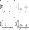
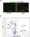
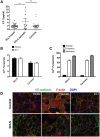
Idiopathic systemic capillary leak syndrome (SCLS) is a rare and potentially fatal vascular disorder characterized by reversible bouts of hypotension and edema resulting from fluid and solute escape into soft tissues. Although spikes in permeability-inducing factors have been linked to acute SCLS flares, whether or not they act on an inherently dysfunctional endothelium is unknown. To assess the contribution of endothelial-intrinsic mechanisms in SCLS, we derived blood-outgrowth endothelial cells (BOEC) from patients and healthy controls and examined gene expression patterns. Ednra, encoding Endothelin receptor A (ETA)-the target of Endothelin 1 (ET-1)-was significantly increased in SCLS BOEC compared to healthy controls. Although vasoconstriction mediated by ET-1 through ETA activation on vascular smooth muscle cells has been well characterized, the expression and function of ETA receptors in endothelial cells (ECs) has not been described. To determine the role of ETA and its ligand ET-1 in SCLS, if any, we examined ET-1 levels in SCLS sera and functional effects of endothelial ETA expression. ETA overexpression in EAhy926 endothelioma cells led to ET-1-induced hyper-permeability through canonical mechanisms. Serum ET-1 levels were elevated in acute SCLS sera compared to remission and healthy control sera, suggesting a possible role for ET-1 and ETA in SCLS pathogenesis. However, although ET-1 alone did not induce hyper-permeability of patient-derived BOEC, an SCLS-related mediator (CXCL10) increased Edrna quantities in BOEC, suggesting a link between SCLS and endothelial ETA expression. These results demonstrate that ET-1 triggers classical mechanisms of vascular barrier dysfunction in ECs through ETA. Further studies of the ET-1-ETA axis in SCLS and in more common plasma leakage syndromes including sepsis and filovirus infection would advance our understanding of vascular integrity mechanisms and potentially uncover new treatment strategies.
Conflict of interest statement
Figures


Similar articles
-
Adrenomedullin surges are linked to acute episodes of the systemic capillary leak syndrome (Clarkson disease).J Leukoc Biol. 2018 Apr;103(4):749-759. doi: 10.1002/JLB.5A0817-324R. Epub 2018 Jan 23. J Leukoc Biol. 2018. PMID: 29360169 Free PMC article.
-
Neutrophil activation in systemic capillary leak syndrome (Clarkson disease).J Cell Mol Med. 2019 Aug;23(8):5119-5127. doi: 10.1111/jcmm.14381. Epub 2019 Jun 18. J Cell Mol Med. 2019. PMID: 31210423 Free PMC article.
-
Vascular endothelial hyperpermeability induces the clinical symptoms of Clarkson disease (the systemic capillary leak syndrome).Blood. 2012 May 3;119(18):4321-32. doi: 10.1182/blood-2011-08-375816. Epub 2012 Mar 12. Blood. 2012. PMID: 22411873 Free PMC article.
-
Raised Serum Levels of Syndecan-1 (CD138), in a Case of Acute Idiopathic Systemic Capillary Leak Syndrome (SCLS) (Clarkson's Disease).Am J Case Rep. 2018 Feb 16;19:176-182. doi: 10.12659/ajcr.906514. Am J Case Rep. 2018. PMID: 29449526 Free PMC article. Review.
-
Endothelin ETA receptor antagonism in cardiovascular disease.Eur J Pharmacol. 2014 Aug 15;737:210-3. doi: 10.1016/j.ejphar.2014.05.046. Epub 2014 Jun 2. Eur J Pharmacol. 2014. PMID: 24952955 Review.
Cited by
-
Developments in the Role of Endothelin-1 in Atherosclerosis: A Potential Therapeutic Target?Am J Hypertens. 2019 Aug 14;32(9):813-815. doi: 10.1093/ajh/hpz091. Am J Hypertens. 2019. PMID: 31145445 Free PMC article. No abstract available.
-
A ligand-independent Tie2-activating antibody reduces vascular leakage in models of Clarkson disease.Sci Adv. 2023 Nov 15;9(46):eadi1394. doi: 10.1126/sciadv.adi1394. Epub 2023 Nov 17. Sci Adv. 2023. PMID: 37976351 Free PMC article.
-
Intrinsic endothelial hyperresponsiveness to inflammatory mediators drives acute episodes in models of Clarkson disease.J Clin Invest. 2024 Mar 19;134(10):e169137. doi: 10.1172/JCI169137. J Clin Invest. 2024. PMID: 38502192 Free PMC article.
-
PARP15 Is a Susceptibility Locus for Clarkson Disease (Monoclonal Gammopathy-Associated Systemic Capillary Leak Syndrome).Arterioscler Thromb Vasc Biol. 2024 Dec;44(12):2628-2646. doi: 10.1161/ATVBAHA.124.321522. Epub 2024 Oct 31. Arterioscler Thromb Vasc Biol. 2024. PMID: 39479769
-
Ultrasound findings and specific intrinsic blood volume expansion therapy for neonatal capillary leak syndrome: report from a multicenter prospective self-control study.Eur J Med Res. 2024 Mar 1;29(1):150. doi: 10.1186/s40001-024-01738-2. Eur J Med Res. 2024. PMID: 38429824 Free PMC article.
References
-
- Clarkson B, Thompson D, Horwith M, Luckey EH (1960) Cyclical edema and shock due to increased capillary permeability. Am J Med 29: 193–216. - PubMed
-
- Lambert M, Launay D, Hachulla E, Morell-Dubois S, Soland V, Queyrel V et al. (2008) High-dose intravenous immunoglobulins dramatically reverse systemic capillary leak syndrome. Crit Care Med 36: 2184–2187. - PubMed
Publication types
MeSH terms
Substances
Grants and funding
LinkOut - more resources
Full Text Sources
Other Literature Sources

