Clinical Trial
. 2015 Jul 15;10(7):e0132011.
doi: 10.1371/journal.pone.0132011. eCollection 2015.

Complete Removal of Extracellular IgG Antibodies in a Randomized Dose-Escalation Phase I Study with the Bacterial Enzyme IdeS--A Novel Therapeutic Opportunity

Affiliations
Clinical Trial

Complete Removal of Extracellular IgG Antibodies in a Randomized Dose-Escalation Phase I Study with the Bacterial Enzyme IdeS--A Novel Therapeutic Opportunity

Lena Winstedt et al. PLoS One. .

Abstract

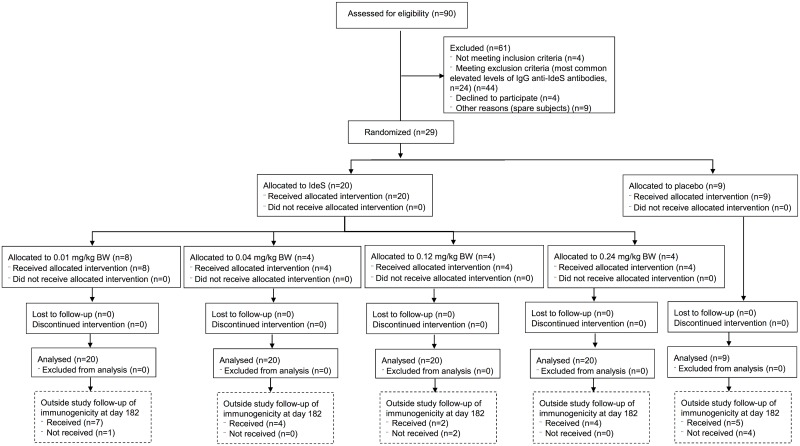
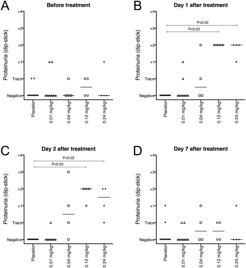
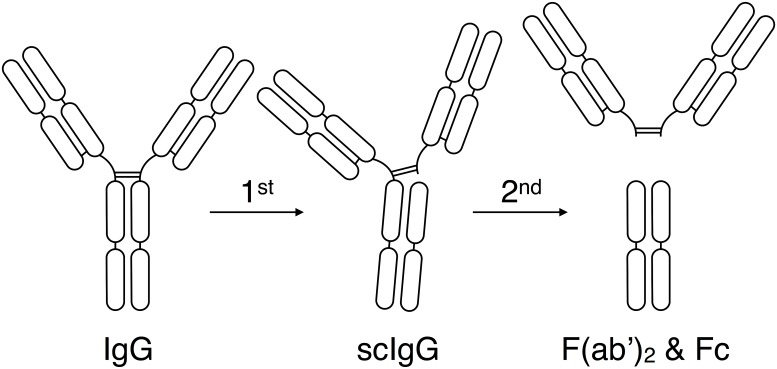
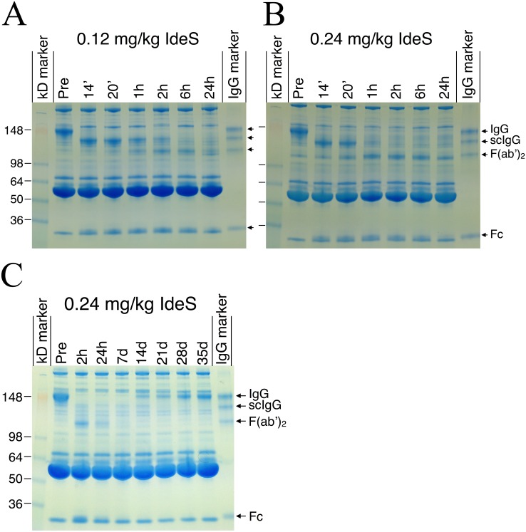
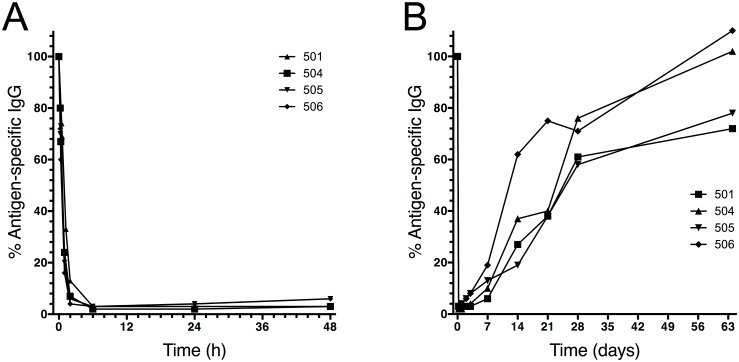
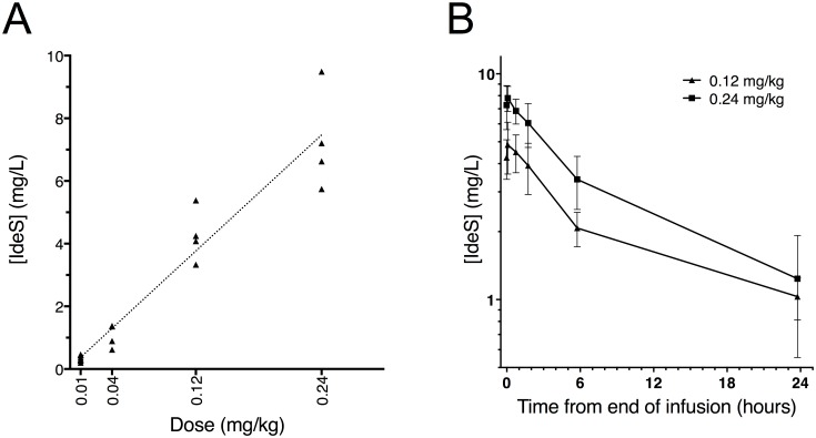
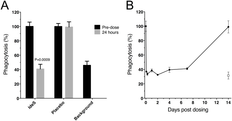
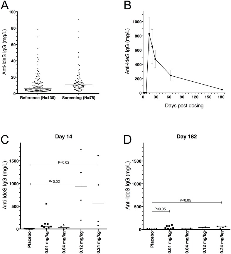
IdeS is a streptococcal protease that cleaves IgG antibodies into F(ab’)2 and Fc fragments with a unique degree of specificity, thereby providing a novel treatment opportunity of IgG-driven autoimmune conditions and antibody mediated transplant rejection. Here we report the results from a first in man, double blinded and randomized study with single ascending doses of IdeS in healthy, male subjects. Twenty healthy subjects were given intravenous single ascending doses of IdeS. With impressive efficacy IdeS cleaved the entire plasma IgG-pool only minutes after dosing. IgG reached nadir 6-24 hours after dosing and then slowly recovered. The half-life of IdeS was 4.9 (±2.8) hours at 0.24 mg/kg with the main fraction eliminated during 24 hours. Already two hours after IdeS-dosing, the phagocytic capacity of IgG/IgG-fragments was reduced to background levels. Importantly, IdeS has the capacity to inactivate Fc-mediated effector function in vivo, was considered safe with no serious adverse events, and without dose limiting toxicity in this study. The complete, rapid, but temporary removal of IgG provides a new potent therapeutic opportunity in IgG-mediated pathogenic conditions.

Trial registration: ClinicalTrials.gov NCT01802697.


Conflict of interest statement

Competing Interests: have read the journal's policy and the authors of this manuscript have the following competing interests: Hansa Medical AB was the formal sponsor and funder of the clinical trial. LW, SJ, EAN, AO, AR, RB, and CK are employed by Hansa Medical AB. LB is an inventor on patent applications related to IdeS and owns shares in Hansa Medical AB. The patent applications are: 1. International publication No: WO/2006/131347 Title: USE OF THE IDES PROTEINASE (FROM S. PYOGENES) FOR TREATING AUTOIMMUNE DISEASES AND GRAFT REJECTION International publication date: 14.12.2006 2. International publication No: WO/2003/051914 Title: IDES, AN IGG-DEGRADING ENZYME OF STREPTOCOCCUS PYOGENES International publication date: 26.06.2003 This does not alter our adherence to PLOS ONE policy on sharing data and materials.

Figures

Fig 1
Fig 1. Participant flow diagram for the safety study on IdeS in healthy volunteers.
The study ended at day 64, but all subjects were asked to come back for additional sampling at day 182 in order to monitor immunogenicity.
Fig 2
Fig 2. Proteinuria was monitored as a safety assessment throughout the study.
Proteinuria was routinely measured at the hospital using Multistix (Siemens) and reported as negative, trace or grade 1–4. Data represents proteinuria in individual subjects at the indicated time-points after treatment. A-C) A mild but significant increase in proteinuria was seen on day 1 (P = 0.0003) and day 2 (P = 0.0002) after treatment with 0.12 and 0.24 mg/kg IdeS. D) On day 7 there were no significant (P = 0.80) differences between groups. The groups (n Placebo = 9, n 0.01 = 8, n 0.04 = 4, n 0.12 = 4 and n 0.24 = 4) were compared using Kruskal-Wallis, One-Way ANOVA. The P-values shown in the graph represent comparisons of the mean rank of each dose-group with the placebo group using Dunn’s Multiple Comparison. The placebo group represents a pool of all subjects treated with placebo from all dose-groups.
Fig 3
Fig 3. Schematic representation of IgG cleavage by IdeS.
Intact human IgG, regardless of isotype, is cleaved by IdeS in two steps. The first step generates a single-cleaved IgG molecule (scIgG) with one intact heavy chain. The second step generates one F(ab’)2 fragment and one homo-dimeric Fc fragment held together by non-covalent interactions.
Fig 4
Fig 4. Qualitative pharmacodynamics analysis by SDS-PAGE showed rapid degradation of IgG.
Non-reducing SDS-PAGE analysis of serum from subjects dosed with A) 0.12 mg/kg BW IdeS and B) 0.24 mg/kg BW IdeS showing protein banding patterns at pre-dosing, 14 min, 20 min, 1, 2, 6 and 24 hours after dosing. C) IgG recovery in serum from one subject in the 0.24 mg/kg BW group at pre-dosing, 2 hours, 24 hours, 7 days, 14, 21, 28 and 35 days after dosing. Arrows to the right in each figure show the different bands in the IgG-marker containing a mix of human IgG, scIgG, F(ab’)2 and Fc. Lines to the left in each figure show the relative molecular mass of the kDa standard. The gels show a representative subject in the 0.12 and 0.24 mg/kg BW IdeS dose groups.
Fig 5
Fig 5. Quantitative pharmacodynamics analysis by ELISA showed rapid degradation of IgG.
Serum IgG levels from all four individual subjects dosed with 0.12 mg/kg BW IdeS (A and B) and all four individual subjects dosed with 0.24 mg/kg BW IdeS (C and D) determined using a validated ELISA method performed by Covance Laboratories Ltd, UK. To be able to follow both early, rapid degradation as well as recovery of IgG, graphs A and C show data up to 48 hours after dosing (x-axis in hours) and graphs B and D show data until last visit (x-axis in days).
Fig 6
Fig 6. Pharmacodynamics of antigen-specific IgG following IdeS treatment.
Human serum samples from the 0.24 mg/kg BW group (n = 4) were addressed for presence of specific IgG against a vaccine mixture of antigens (diphtheria, pertussis, tetanus, polio and Haemophilus influenzae type b). The results are given as percent remaining IgG on the y-axis compared to the start value for each subject. To be able to follow both early, rapid degradation as well as recovery of IgG, graph A shows data up to 48 hours after dosing (x-axis in hours) and graph B shows data until last visit (x-axis in days).
Fig 7
Fig 7. Pharmacokinetics of IdeS in serum.
IdeS concentrations in serum samples from study subjects were determined by selected reaction monitoring mass spectrometry targeting four IdeS specific tryptic peptides. A) Comparison of serum IdeS concentration one minute before end of infusion versus dose levels of IdeS (0.01, 0.04, 0.12, and 0.24 mg/kg BW). Individual mean of two to four peptides. B) Comparison of serum concentration of mean values of two to four peptides versus time profiles up to 24 hours after infusion of 0.12 or 0.24 mg/kg BW IdeS (n = 8). Mean ± SEM.
Fig 8
Fig 8. Serum from subjects dosed with IdeS showed impaired phagocytosis capacity.
The opsonizing capacity of IgG in human serum was measured as percent of effector cells with at least one engulfed fluorescent serum-coated bead. Effector cells were gated and bead uptake was monitored as percent of cells shifted in the FL2 channel. Due to the extreme brightness of the fluorescent beads the signal could not me measured within the dynamic range of the FL1 channel and were instead monitored in FL2. A) Before and 24 hours after dosing of 0.24 mg/kg BW IdeS vs. placebo treated subjects. Pre-dose phagocytosis level for each individual was set to 100% and background is spontaneous uptake of beads in the absence of serum, n = 4 in the IdeS group and n = 2 in the placebo group. Mean of double wells/subject ± SD are shown. P-value was calculated using Mann-Whitney. B) Kinetics of the phagocytic potential in serum is shown for one representative subject in the 0.24 mg/kg BW group at different time-points (pre-dose, 2, 6, 24, 48 hours, 4, 7 and 14 days). Pre-dose phagocytosis level was set to 100% and background is spontaneous uptake of beads in the absence of serum (open box). Mean of double wells ± SD for this subject.
Fig 9
Fig 9. Anti-IdeS antibodies were followed before and throughout the study.
Human serum samples were analyzed using the IdeS-ImmunoCAP (Thermo Fisher Scientific) on a Phadia 250 instrument. The cut off (LLOQ) for IgG was 2 mg/L. A) Samples from 130 human donors (reference) were compared to the 78 healthy human male subjects screened in this study (screening). The lines show median for the reference group (6.1 mg/L) and the screening group (10.6 mg/L). B) Kinetics of the anti-IdeS IgG levels shown as a mean for the 0.12 and 0.24 mg/kg BW groups (n = 8; mean ± SEM). No increase in anti-IdeS IgG is seen in any of the subjects prior to day 14. C) Anti-IdeS IgG levels shown for the separate groups at day 14 (n Placebo = 9, n 0.01 = 8, n 0.04 = 4, n 0.12 = 4 and n 0.24 = 4), and D) at day 182 (n Placebo = 5, n 0.01 = 7, n 0.04 = 4, n 0.12 = 2 and n 0.24 = 4). The subjects were asked to come back for an outside the study protocol sample on day 182. 17 out of 20 on active and 5 out of 9 on placebo volunteered. The lines show median level for each group. In C and D the groups were compared using Kruskal-Wallis, One-Way ANOVA, P = 0.0003 and P = 0.0082 respectively. The P-values shown in the graph represent comparisons of the mean rank of each dose-group with the placebo group using Dunn’s Multiple Comparison. The placebo group contains all subjects from all dose-groups treated with placebo.

Similar articles

Cited by

References

    1. von Pawel-Rammingen U, Johansson BP, Björck L (2002) IdeS, a novel streptococcal cysteine proteinase with unique specificity for immunoglobulin G. EMBO J 21: 1607–1615. - PMC - PubMed
    1. Carapetis JR, Steer AC, Mulholland EK, Weber M (2005) The global burden of group A streptococcal diseases. Lancet Infect Dis 5: 685–694. - PubMed
    1. Wenig K, Chatwell L, von Pawel-Rammingen U, Björck L, Huber R, Sondermann P (2004) Structure of the streptococcal endopeptidase IdeS, a cysteine proteinase with strict specificity for IgG. Proc Natl Acad Sci U S A 101: 17371–17376. - PMC - PubMed
    1. Vincents B, von Pawel-Rammingen U, Björck L, Abrahamson M (2004) Enzymatic characterization of the streptococcal endopeptidase, IdeS, reveals that it is a cysteine protease with strict specificity for IgG cleavage due to exosite binding. Biochemistry 43: 15540–15549. - PubMed
    1. Johansson BP, Shannon O, Björck L (2008) IdeS: a bacterial proteolytic enzyme with therapeutic potential. PLoS One 3: e1692 10.1371/journal.pone.0001692 - DOI - PMC - PubMed

Publication types

MeSH terms

Associated data