. 2016 Feb;117(2):351-60.
doi: 10.1002/jcb.25277.

Multiple Levels of Regulation of Sororin by Cdk1 and Aurora B

Affiliations

Multiple Levels of Regulation of Sororin by Cdk1 and Aurora B

Michael T Borton et al. J Cell Biochem. 2016 Feb.

Abstract

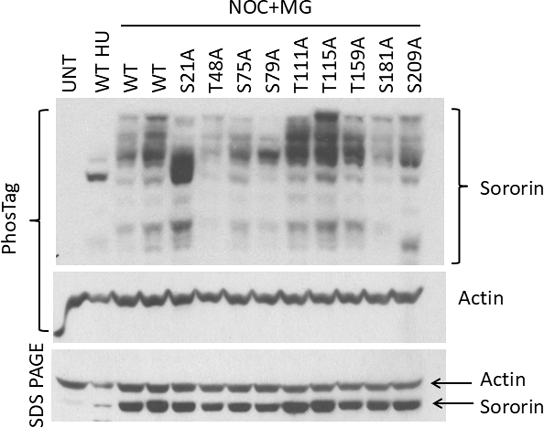
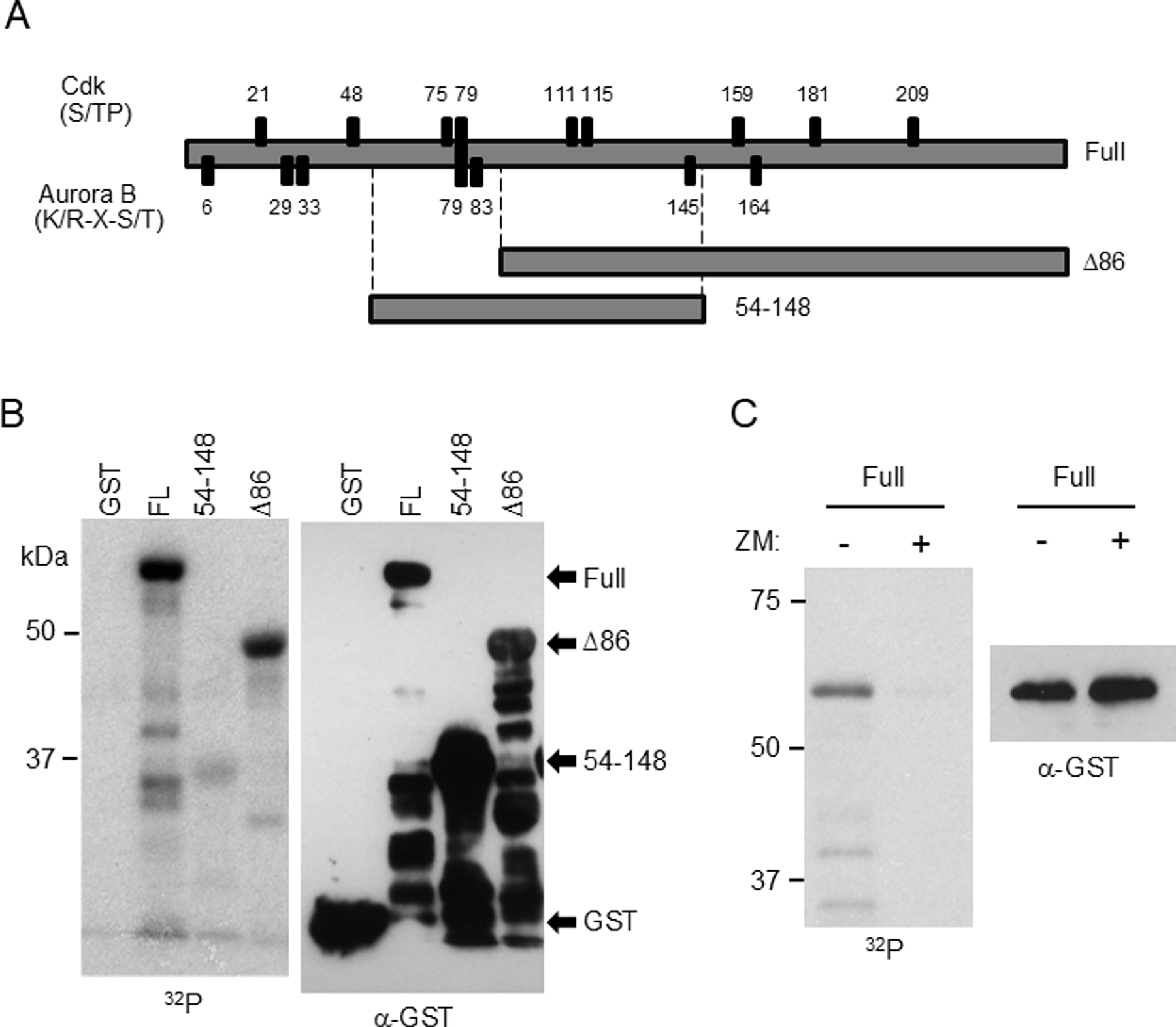
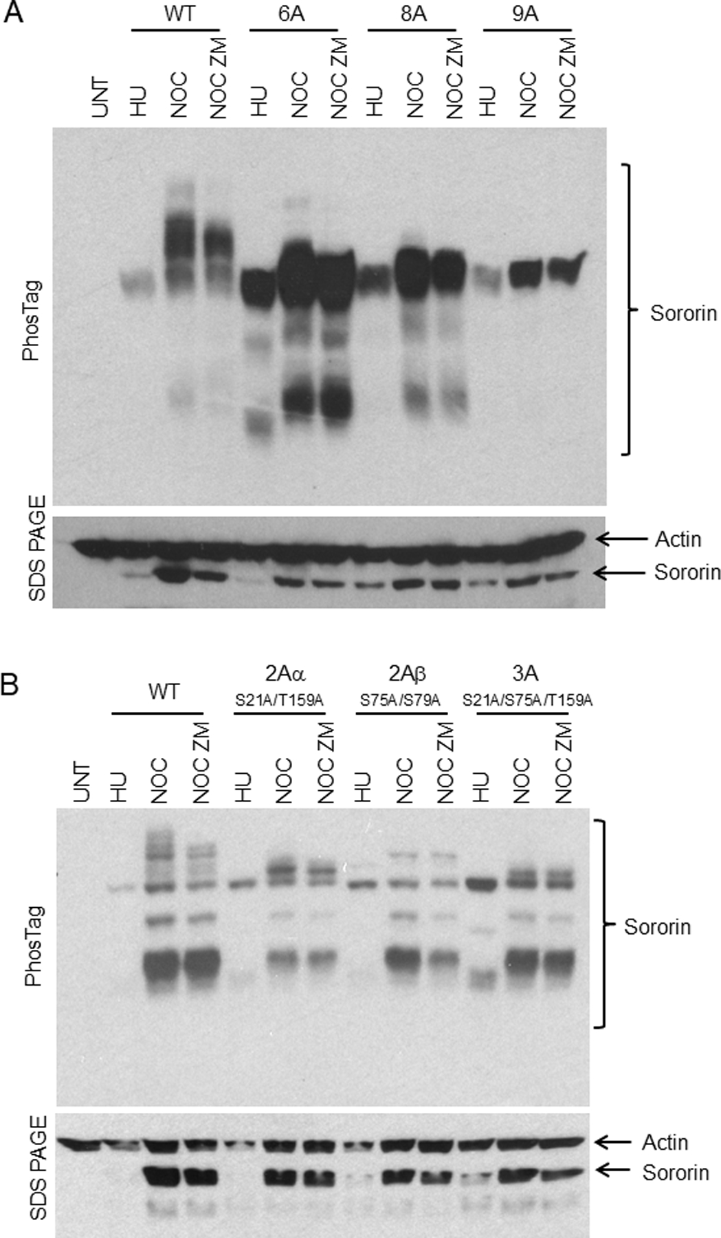
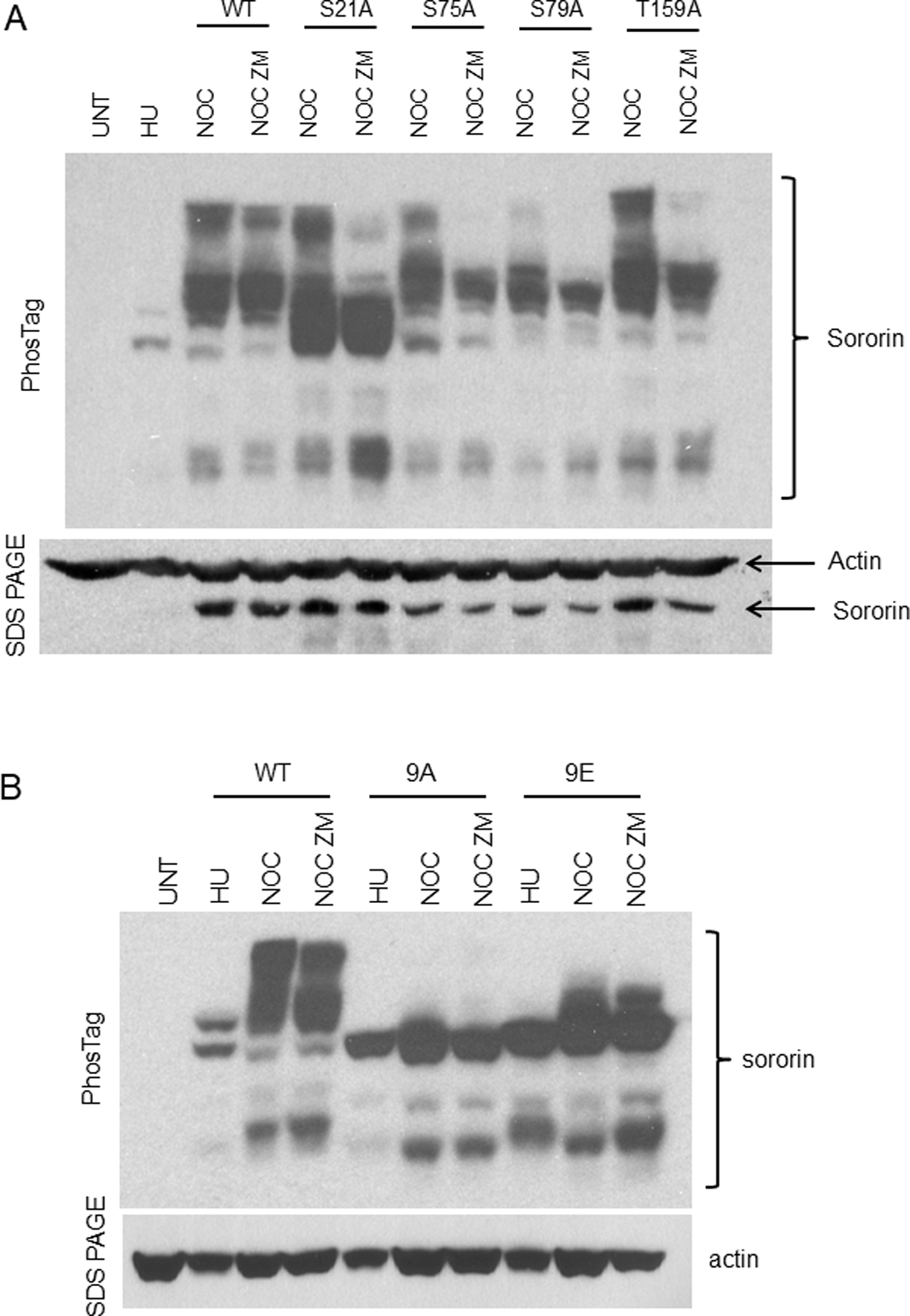
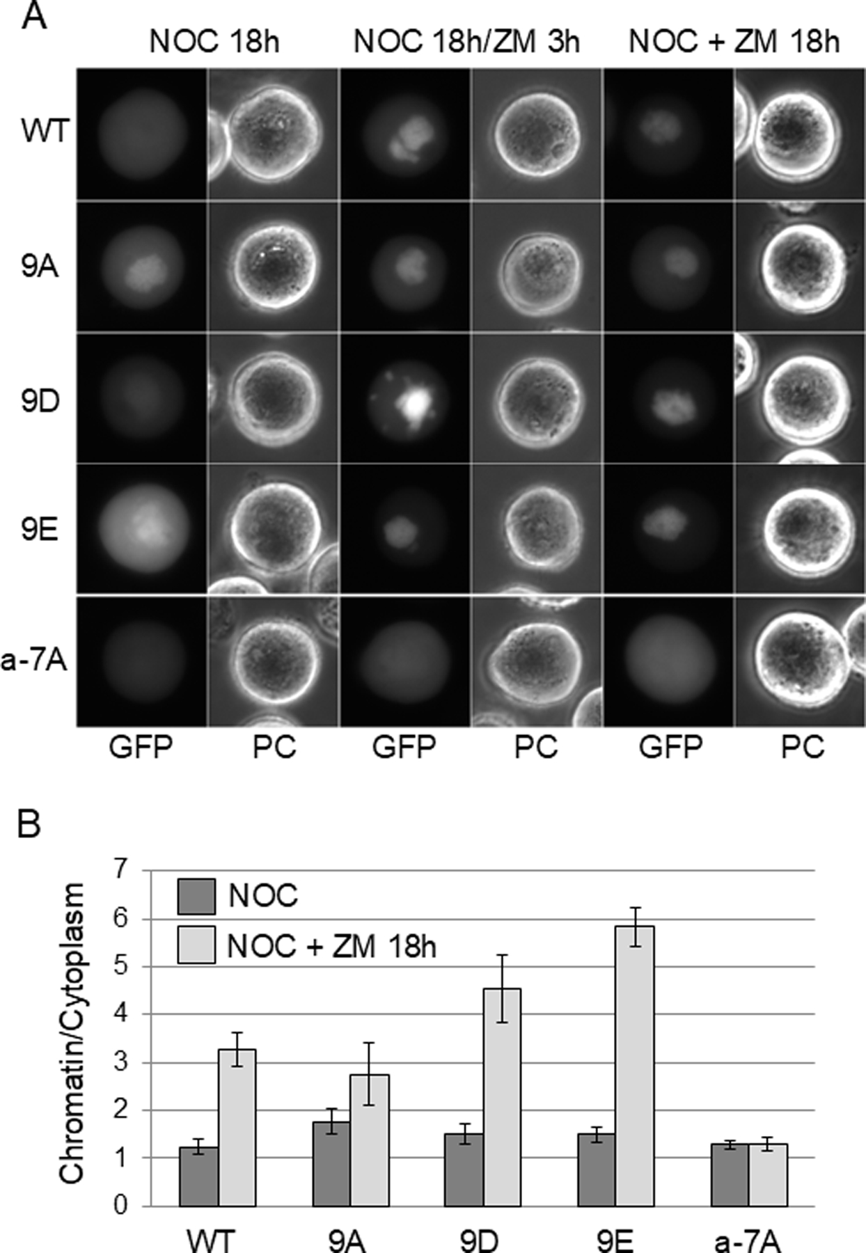
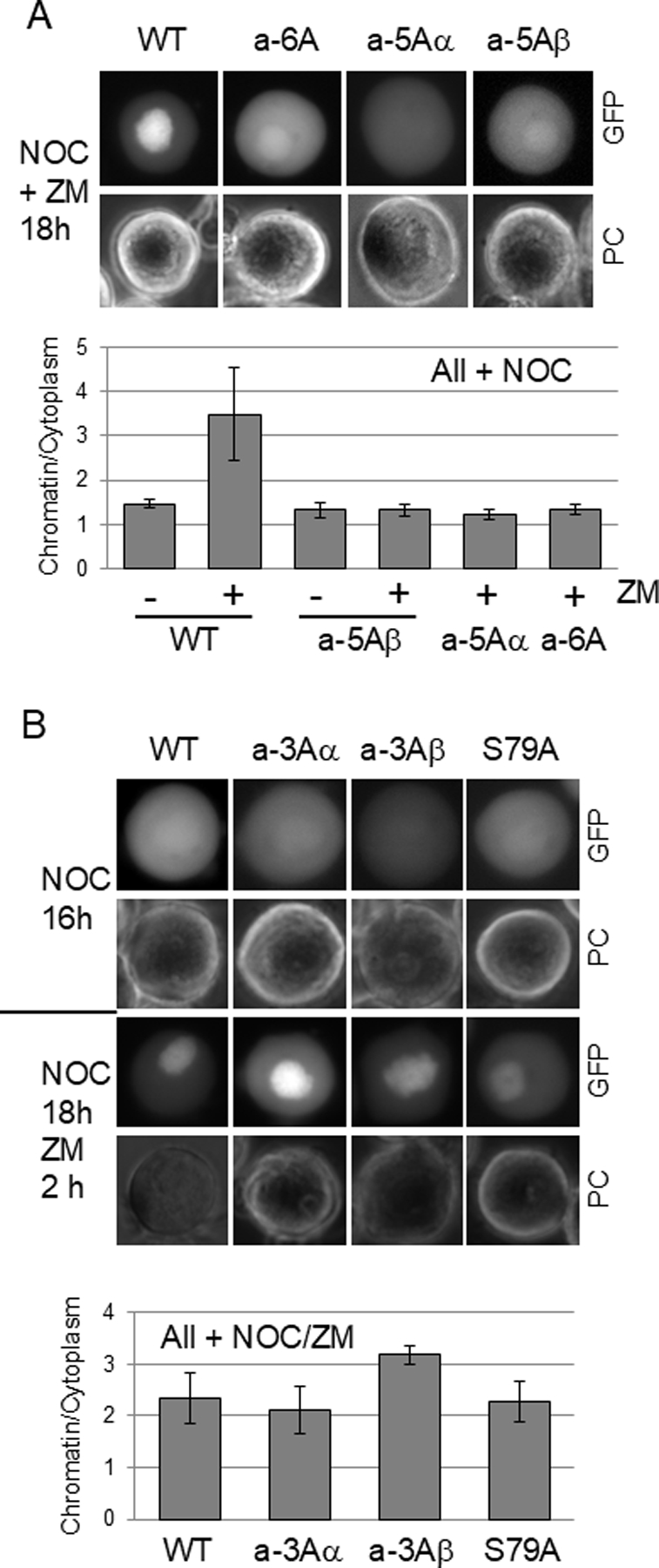
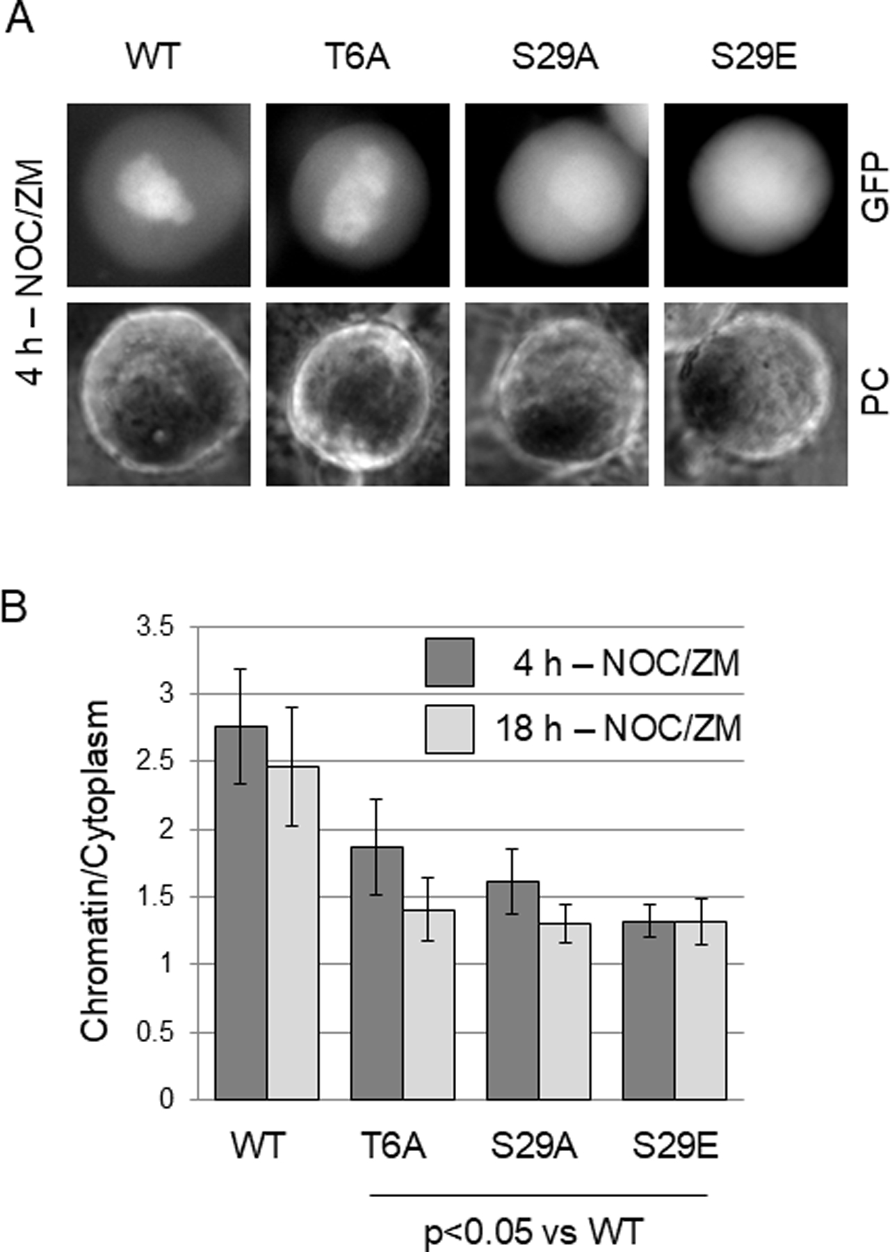
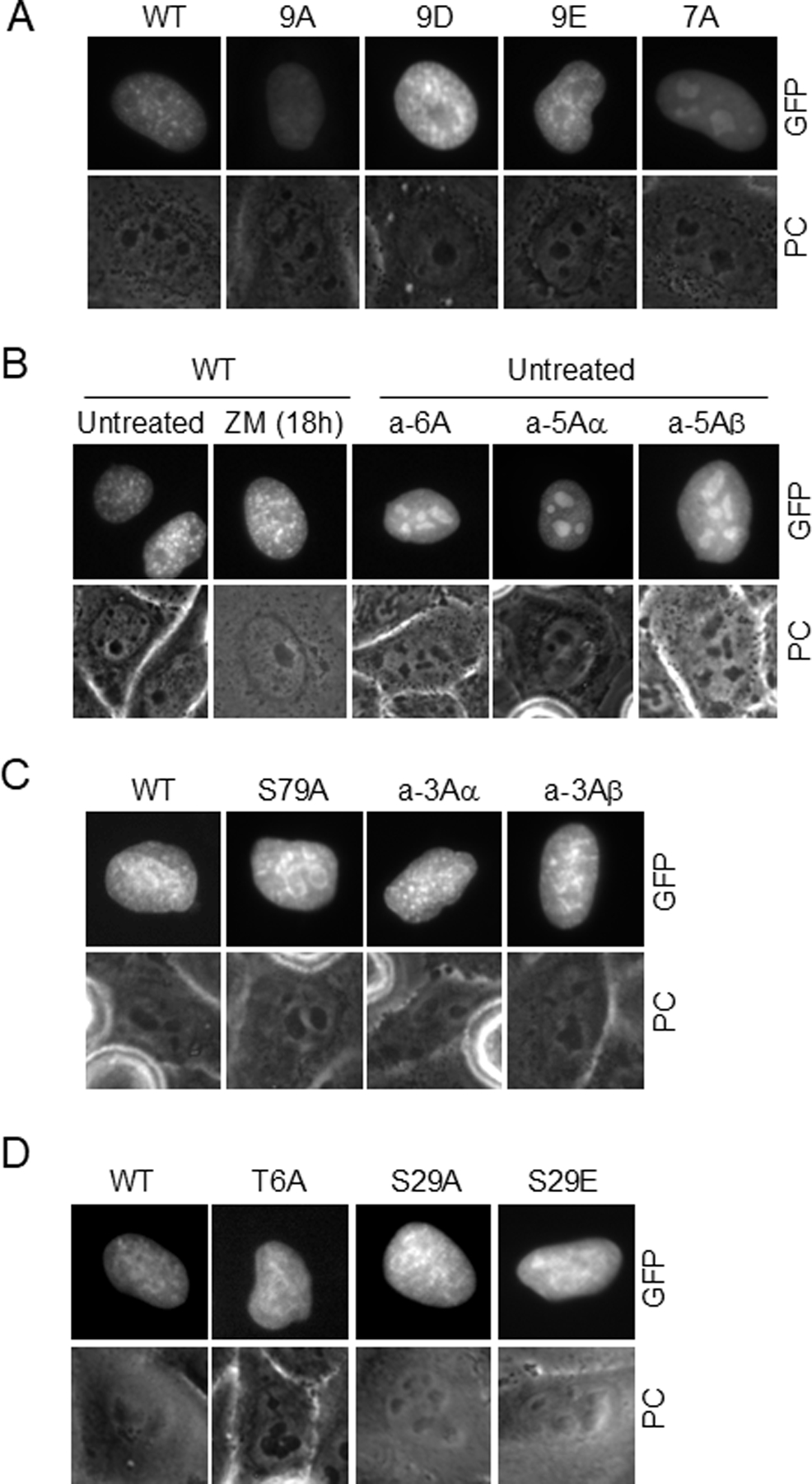
The cohesin complex holds sister chromatids together until all chromosomes are properly attached to the mitotic spindle. Cleavage of the cohesin subunit Rad21 at the metaphase to anaphase transition allows separation of sister chromatids and is fundamental for the creation of identical daughter cells. Sororin blocks removal of cohesin from chromosomes from S phase until mitosis. In mitosis, Sororin is phosphorylated by Cdk1 releasing it from the cohesin complex. Aurora B phosphorylation of Sororin may play a similar role as Cdk1. Using PhosTag electrophoresis, we detect multiple Sororin species suggesting that phosphorylation of Sororin in mitosis is heterogeneous. Mutating the Cdk1 consensus site S21 to alanine eliminates many of the phosphorylated species suggesting that S21 is a key site of phosphorylation in vivo. Inhibiting Aurora B reduces phosphorylation of Sororin in cells, but only if Cdk1 sites are intact suggesting that some phosphorylation events on Sororin may be sequential. Surprisingly, mutating Aurora B consensus sites in Sororin causes it to relocalize to the nucleolus during interphase and blocks its interaction with chromosomes in Aurora B-inhibited cells. These observations indicate that phosphorylation plays unexpected roles in regulating the subcellular localization of Sororin.

Keywords: COHESIN; MITOSIS; NUCLEOLUS; PHOSPHORYLATION; PHOSTAG.


Conflict of interest statement

Conflict of Interest

The authors state that they have no conflict of interest to declare.

Figures

Figure 1:
Figure 1:
PhosTag analysis of Cdk1 site mutants of Sororin. HeLa M cells transiently transfected with V5-tagged Sororin mutants were either blocked in S phase with hydroxyurea (HU) or in mitosis with nocodazole (NOC) for 18 hours. MG132 (MG) was added for an additional two hours to allow for comparison to inhibitor-treated samples in Figures 3–5. Two separate wild-type (WT)-transfected lysates were run to determine variability. In the top panel, lysates were separated by PhosTag electrophoresis and probed with an antibody to Sororin, followed by stripping and reprobing for Actin. A smaller aliquot of the same lysates was separated by regular SDS-PAGE for comparison. “UNT” : untransfected.
Figure 2:
Figure 2:
Phosphorylation of Sororin by Aurora B in vitro. GST-tagged Sororin fragments were purified from E. coli and incubated with recombinant Aurora B and the IN-box of INCENP in the presence of 32P-labeled ATP. (A) Schematic showing Cdk and Aurora B consensus sites as well as regions represented in the Sororin fragments used as substrates. (B). Autoradiograph of in vitro-phosphorylated Sororin is shown in the left panel. The same blot, probed with an antibody to GST is shown on the right. (C) ZM447439 added to in vitro reactions to determine specificity.
Figure 3:
Figure 3:
PhosTag analysis of Sororin after inhibition of Aurora B. HeLa M cells were transiently transfected with the indicated compound mutants of Sororin. Cells were treated for 18 hours with either hydroxyurea (HU) to block them in S phase or with nocodazole to block them in mitosis. The nocodazole-treated samples were then treated with MG132 alone (“NOC”) or MG132 + 2.5μM ZM447439 (“NOC ZM”) for 2 hours. ZM447439 is added to inhibit Aurora B, and MG132 is added to prevent mitotic exit when Aurora B is inhibited. In the top panel, lysates were separated by PhosTag electrophoresis before Western analysis. In the bottom panel, an aliquot of the same lysate was separated by standard SDS-PAGE and probed with antibodies to Sororin and Actin for loading. Experiments shown in “A and B” were carried out under the same conditions with different mutants transfected as indicated. “UNT”: untransfected.
Figure 4:
Figure 4:
PhosTag analysis of single Cdk1 site mutants of Sororin after inhibition of Aurora B. HeLa M transfection and PhosTag analysis was carried out as described in Figure 3. (A) Single site mutations at Cdk1 consensus sites incorporated into V5-tagged Sororin. (B) Comparison of Sororin-9A and Sororin-9E by PhosTag.
Figure 5:
Figure 5:
Localization of wild-type and mutant Sororin-GFP in response to Aurora B inhibition. HeLa M cells were transiently transfected with Sororin-GFP. Live cells were imaged by wide-field fluorescence microscopy. (A) Transfected cells were exposed to nocodazole (NOC) for 15 hours followed by ZM447439 (ZM) for an additional 3 hours (NOC 18h/ZM 3h). In the columns on the right, cells were exposed to nocodazole and ZM447439 for 18 hours (NOC + ZM 18h). ZM447439 for either 3 or 18 hours causes association of Sororin with chromatin except in the mutant a-7A which stays dispersed in the mitotic cytoplasm. “PC”: Phase contrast. (B) Quantitation of the chromatin/cytoplasm ratio in images of live cells. Pixel intensity in the central and peripheral regions of the cell were determined and are shown as a ratio. Bars represent standard deviation.
Figure 6:
Figure 6:
Localization of additional mutants of Sororin after inhibition of Aurora B. HeLa M cells were transfected with mutants of Sororin-GFP in which various Aurora B consensus sites were converted to alanine and then analyzed by fluorescence microscopy. (A) Sororin mutants exposed to nocodazole and ZM447439 for 18 hours. (B) Sororin mutants arrested in mitosis with nocodazole and imaged either before (NOC 16h) or after a 2 hour treatment with ZM447439 (NOC 18h ZM 2h). For both “A” and “B”, the chromatin/cytoplasm expression ratio is shown below each corresponding panel.
Figure 7:
Figure 7:
Effect of mutations at T6 and S29 on the localization of Sororin-GFP after Aurora B inhibition. Live HeLa M cells transiently transfected with the indicated mutants were analyzed by wide-field fluorescence microscopy. (A) Representative images of Sororin-GFP transfected cells after inhibition of Aurora B. One day after transfection, nocodazole and ZM447439 were added simultaneously. Images of live cells were captured 4 hours later. (B) Quantitation of chromatin/cytoplasm ratio. One day after transfection, nocodazole and ZM447439 were added simultaneously. Cells were imaged either 4 or 18 hours later.
Figure 8:
Figure 8:
Analysis of phosphorylation sites required to remove Sororin-GFP from the nucleolus. HeLa M cells were transiently transfected with the indicated mutants and live interphase cells imaged by wide-field fluorescence microscopy. (A) Interphase nuclei in untreated cultures indicate dispersed, but punctate staining in WT, 9A, 9D, 9E. Mutant a-7A shows nucleoloar enrichment. (B) Comparison of Sororin mutants bearing either 5 or 6 Aurora consensus site mutants to the wild-type protein. (C) Comparison of wild-type, S79A or two triple Sororin mutants exposed to nocodazole for 18 hours. Cells that had not yet entered mitosis were imaged indicating a wild-type localization in these mutants. Similar results were obtained in untreated cells. (D) Effect of mutations at T6 and S29 on interphase Sororin localization. Cells were untreated at the time of imaging.

Similar articles

Cited by

References

    1. Barber TD, McManus K, Yuen KW, Reis M, Parmigiani G, Shen D, Barrett I, Nouhi Y, Spencer F, Markowitz S, Velculescu VE, Kinzler KW, Vogelstein B, Lengauer C, Hieter P. 2008. Chromatid cohesion defects may underlie chromosome instability in human colorectal cancers. Proc Natl Acad Sci U S A 105:3443–8. - PMC - PubMed
    1. Beausoleil SA, Villen J, Gerber SA, Rush J, Gygi SP. 2006. A probability-based approach for high-throughput protein phosphorylation analysis and site localization. Nat Biotechnol 24:1285–92. - PubMed
    1. Cantin GT, Yi W, Lu B, Park SK, Xu T, Lee JD, Yates JR 3rd. 2008. Combining protein-based IMAC, peptide-based IMAC, and MudPIT for efficient phosphoproteomic analysis. J Proteome Res 7:1346–51. - PubMed
    1. Chen RQ, Yang QK, Lu BW, Yi W, Cantin G, Chen YL, Fearns C, Yates JR 3rd, Lee JD. 2009. CDC25B mediates rapamycin-induced oncogenic responses in cancer cells. Cancer Res 69:2663–8. - PMC - PubMed
    1. Dephoure N, Zhou C, Villen J, Beausoleil SA, Bakalarski CE, Elledge SJ, Gygi SP. 2008. A quantitative atlas of mitotic phosphorylation. Proc Natl Acad Sci U S A 105:10762–7. - PMC - PubMed

Publication types

LinkOut - more resources