. 2015 Jul 17:8:40.
doi: 10.1186/s12920-015-0118-9.

Integrated sequence and expression analysis of ovarian cancer structural variants underscores the importance of gene fusion regulation

Affiliations

Integrated sequence and expression analysis of ovarian cancer structural variants underscores the importance of gene fusion regulation

Vinay K Mittal et al. BMC Med Genomics. .

Abstract

Background: Genomic rearrangements or structural variants (SVs) are one of the most common classes of mutations in cancer.

Methods: An integrated DNA sequencing and transcriptional profiling (RNA sequence and microarray gene expression data) analysis was performed on six ovarian cancer patient samples. Matched sets of control (whole blood) samples from these same patients were used to distinguish cancer SVs of germline origin from those arising somatically in the cancer cell lineage.

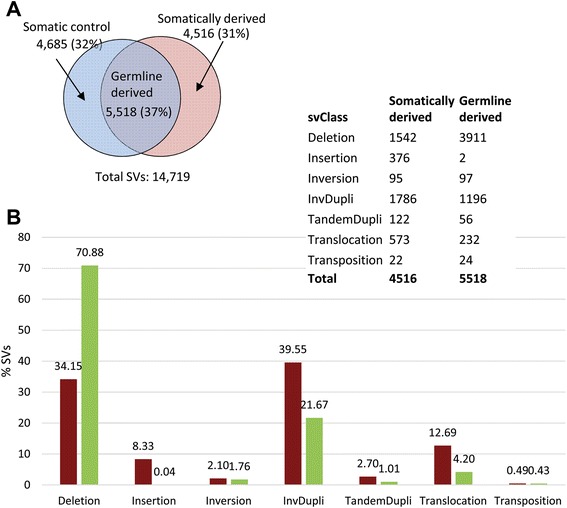
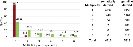
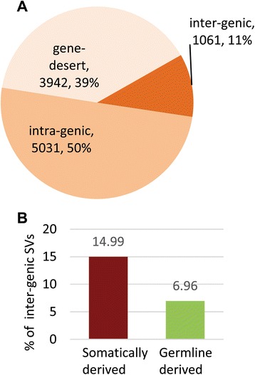
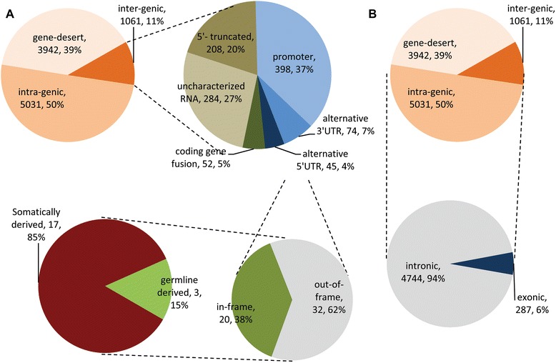
Results: We detected 10,034 ovarian cancer SVs (5518 germline derived; 4516 somatically derived) at base-pair level resolution. Only 11 % of these variants were shown to have the potential to form gene fusions and, of these, less than 20 % were detected at the transcriptional level.

Conclusions: Collectively our results are consistent with the view that gene fusions and other SVs can be significant factors in the onset and progression of ovarian cancer. The results further indicate that it may not only be the occurrence of these variants in cancer but their regulation that contributes to their biological and clinical significance.


Figures

Fig. 1
Fig. 1
a The number (percentage) of germline and somatically derived ovarian cancer SVs in six ovarian cancer patient samples. Circles represent the total number of SVs detected in somatic control (whole blood; blue circle) and cancer (red circle) tissue samples collected from six ovarian cancer patients. SVs identified in both the control and cancer samples were classified as germline derived, while SVs detected in only the cancer samples were classified as somatically derived. b Distribution of SVs across structural categories. Somatically derived (red) and germline derived (green) SVs were further categorized according to the underlying genomic rearrangement. Deletions were the most abundant category accounting for the majority (~71 %; see percentages on top of bars) of the germline derived SVs. Corresponding data are shown in the Table (top right corner). InvDupli: inverted-duplications, TandemDupli: tandem-duplication
Fig. 2
Fig. 2
Multiplicity of SVs across samples. The X-axis represents multiplicity (number of occurrences) of somatically derived (red bars) and germline derived (green bars) SVs across cancer samples. The Y-axis represents percentage of SVs present in a particular multiplicity. The table presents the numerical distribution of SVs across the cancer samples
Fig. 3
Fig. 3
Classification of ovarian cancer SVs based on breakpoints. SVs are depicted by black-grey boxes, reference transcripts are represented by the blue and green boxes. Thick boxes represent open-reading-frame or coding sequences (CDS) while thin boxes represent UTRs. a Intergenic SVs – breakpoints map to annotated genes located at distant genomic locations; (b) intragenic SVs- breakpoints map within the same gene; (c) gene desert- breakpoints map to un-annotated genomic regions (gene deserts)
Fig. 4
Fig. 4
Distribution of SVs across classes. a Distribution of total detected SVs among classes. b Distribution of intergenic SVs between somatically derived and germline derived SVs. Intergenic SVs are significantly enriched (p-value < 0.05) for somatically derived variants
Fig. 5
Fig. 5
Distribution of intergenic and intragenic ovarian cancer SVs. a Distribution of germline and somatically derived intergenic SVs; (b) Distribution of intronic and exonic intragenic SVs
Fig. 6
Fig. 6
Structure of six intergenic SVs resulting in “in-frame” coding gene fusions. The figure represents the structure of the gene fusion and associated protein domains. Square boxes with numbers represent exons (5′ partner gene: blue, 3′ partner gene: orange), red lines represent the fusion breakpoint, gene symbols and corresponding chromosomes (in parenthesis) are shown on top of each gene fusion structure)

Similar articles

Cited by

References

    1. Lupski JR, Stankiewicz P. Genomic disorders: molecular mechanisms for rearrangements and conveyed phenotypes. PLoS Genet. 2005;1:e49. doi: 10.1371/journal.pgen.0010049. - DOI - PMC - PubMed
    1. Pleasance ED, Cheetham RK, Stephens PJ, McBride DJ, Humphray SJ, Greenman CD, Varela I, Lin ML, Ordonez GR, Bignell GR, Ye K, Allpaz J, Bauer MJ, Beare D, Butler A, Carter RJ, Chen L, Cox AJ, Edkins S, Kokko-Gonzales PI, Gormley NA, Grocock RJ, Haudenschild CD, Hims MM, James T, Jia M, Kingsbury Z, Leroy C, Marshall J, Menzies A, Mudie LJ, Ning Z, Royce T, Schulz-Trieglaff OB, Spiridou A, Stebbins LA, Szajkowski L, Teague J, Williamson D, Chin L, Ross MT, Campbell P, Bentley DR, Futreal PA, Stratton MR. A comprehensive catalogue of somatic mutations from a human cancer genome. Nature. 2010;463:191–196. doi: 10.1038/nature08658. - DOI - PMC - PubMed
    1. Futreal PA, Coin L, Marshall M, Down T, Hubbard T, Wooster R, Rahman N, Stratton MR. A census of human cancer genes. Nat Rev Can. 2004;4:177–183. doi: 10.1038/nrc1299. - DOI - PMC - PubMed
    1. Stratton MR, Campbell PJ, Futreal PA. The cancer genome. Nature. 2009;458:719–724. doi: 10.1038/nature07943. - DOI - PMC - PubMed
    1. Edwards PA. Fusion genes and chromosome translocations in the common epithelial cancers. J Path. 2010;220:244–254. - PubMed