. 2015 Jul 16:13:32.
doi: 10.1186/s12964-015-0112-z.

Orai/CRACM1 and KCa3.1 ion channels interact in the human lung mast cell plasma membrane

Affiliations

Orai/CRACM1 and KCa3.1 ion channels interact in the human lung mast cell plasma membrane

S Mark Duffy et al. Cell Commun Signal. .

Abstract

Background: Orai/CRACM1 ion channels provide the major Ca(2+) influx pathway for FcεRI-dependent human lung mast cell (HLMC) mediator release. The Ca(2+)-activated K(+) channel KCa3.1 modulates Ca(2+) influx and the secretory response through hyperpolarisation of the plasma membrane. We hypothesised that there is a close functional and spatiotemporal interaction between these Ca(2+)- and K(+)-selective channels.

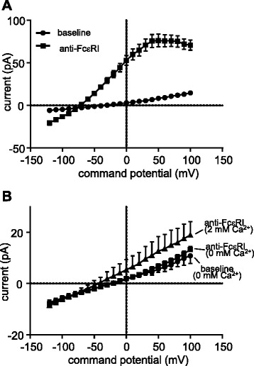
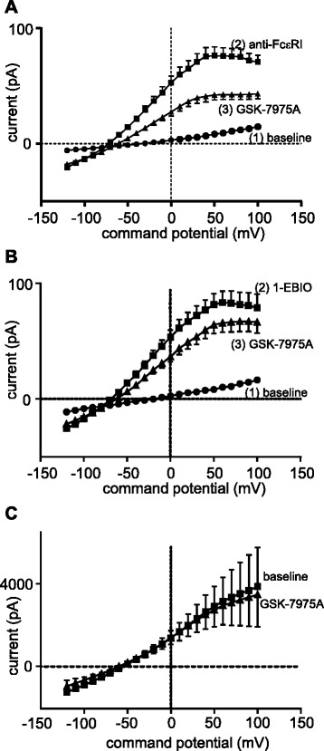
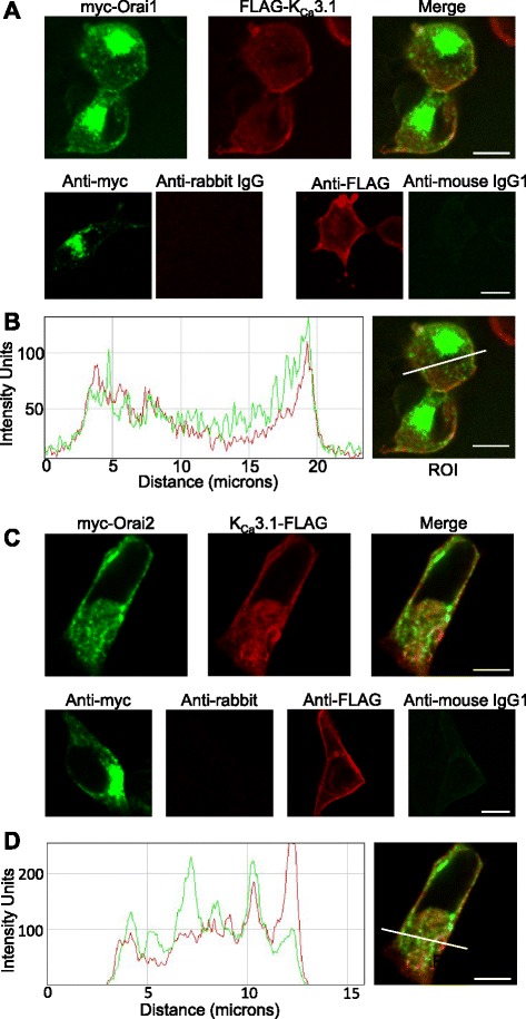
Results: Activation of FcεRI-dependent HLMC KCa3.1 currents was dependent on the presence of extracellular Ca(2+), and attenuated in the presence of the selective Orai blocker GSK-7975A. Currents elicited by the KCa3.1 opener 1-EBIO were also attenuated by GSK-7975A. The Orai1 E106Q dominant-negative mutant ablated 1-EBIO and FcεRI-dependent KCa3.1 currents in HLMCs. Orai1 but not Orai2 was shown to co-immunoprecipitate with KCa3.1 when overexpressed in HEK293 cells, and Orai1 and KCa3.1 were seen to co-localise in the HEK293 plasma membrane using confocal microscopy.

Conclusion: KCa3.1 activation in HLMCs is highly dependent on Ca(2+) influx through Orai1 channels, mediated via a close spatiotemporal interaction between the two channels.


Figures

Fig. 1
Fig. 1
FcεRI-dependent HLMC KCa3.1 currents require the presence of external Ca2+. a Current–voltage curves from HLMCs activated via FcεRI in the presence external Ca2+ (2 mM). b Current–voltage curves from HLMCs activated via FcεRI in the absence of external Ca2+. Typical KCa3.1 currents appear in the presence of Ca2+ (2 mM) but not when it is absent. Small KCa3.1 currents appear in (b) when Ca2+ is then introduced. Data presented as mean ± SEM
Fig. 2
Fig. 2
FcεRI- and 1-EBIO-dependent HLMC KCa3.1 currents are attenuated by an Orai channel blocker. a KCa3.1 currents induced following FcεRI-dependent activation are attenuated in HLMCs following addition of the Orai channel blocker GSK-7975A (n = 27 cells). b KCa3.1 currents induced following 1-EBIO-dependent activation are attenuated following addition of the Orai channel blocker GSK-7975A (n = 29 cells). Data presented as mean ± SEM. c Overexpressed KCa3.1 channels in HLMCs were constitutively active and were not blocked by GSK-7975A (1 μM)(n = 4 cells, p = 0.43 at +40 mV)
Fig. 3
Fig. 3
FcεRI- and 1-EBIO-dependent HLMC KCa3.1 currents are inhibited by expression of an Orai1-E106Q dominant-negative mutant. Transduction of HLMCs with an Orai1-E106Q dominant-negative mutant ablated (a) 1-EBIO- dependent and (b) FcεRI-dependent KCa3.1 currents. For clarity, data are presented as the subtracted net activation-dependent currents (activation minus baseline) for each condition, expressed as mean ± SEM
Fig. 4
Fig. 4
Orai1 and KCa3.1 proteins co-immunoprecipitate. a Western blots using either an antibody recognising the myc epitope (left) or an antibody recognising the FLAG epitope (right) of HEK293 cell lysates. Lysates expressed either myc epitope tagged Orai1, FLAG epitope-tagged KCa3.1, or both as indicated in the panel above. b Lysates of HEK293 cells expressing the indicated proteins were immunoprecipitated with an anti-myc antibody. Immunoprecipitates were then Western blotted using either an anti-Orai1 antibody (left) or an anti-FLAG antibody (right). c As (b) except cell lysates were immunoprecipitated with an anti-FLAG antibody and then Western blotted with an anti-FLAG antibody (left) or an anti-myc antibody (right). Blots shown are representative of 3 independent experiments
Fig. 5
Fig. 5
Orai2 and KCa3.1 proteins do not co-immunoprecipitate under the conditions used to co-immunoprecipitate Orai1 and KCa3.1. a Western blots using either an antibody recognising the myc epitope (left) or an antibody recognising the FLAG epitope (right) of HEK293 cell lysates. Lysates expressed either myc epitope tagged Orai2, FLAG epitope-tagged KCa3.1, or both as indicated in the panel above. b HEK293 cell lysates expressing the proteins indicated in the panel above were immunoprecipitated with an anti-myc antibody. Immunoprecipitates were then Western blotted using either an anti-Orai2 antibody (left) or an anti-FLAG antibody (right). Control HEK293 cell lysate expressing KCa3.1-FLAG protein. c As (b) except cell lysates were immunoprecipitated with an anti-FLAG antibody and then Western blotted with an anti-Orai2 antibody (left) or an anti-FLAG antibody (right). Control HEK293 cell lysate expressing Orai2-myc protein. Blots shown are representative of 3 independent experiments
Fig. 6
Fig. 6
Orai1 and KCa3.1 co-localise in the plasma membrane. a HEK293 cells, dually transfected with FLAG-tagged KCa3.1 and myc-tagged Orai1 and then immunostained, show co-localisation in the plasma membrane by single plane confocal microscopy (top panels). Dually transfected HEK293 show negative staining for appropriate isotype controls (bottom panels): rabbit IgG control, dual stained with anti-myc, and mouse IgG1 control dual stained with anti-FLAG. b Fluorescence intensity plot shows increased fluorescence at the plasma membrane. myc-Orai1 is shown in green and FLAG-KCa3.1 in red. Arrows indicate increased fluorescence where the region of interest (ROI) intersects the plasma membrane. c HEK293 cells, dually transfected with FLAG-tagged KCa3.1 and myc-tagged Orai2 and then immunostained, show poor co-localisation in the plasma membrane by single plane confocal microscopy (top panels). Dually transfected HEK293 show negative staining for appropriate isotype controls (bottom panels): rabbit IgG control, dual stained with anti-myc, and mouse IgG1 control dual stained with anti-FLAG. d Fluorescence intensity plot shows poor co-localisation of KCa3.1 and Orai2 signals. myc-Orai2 is shown in green and FLAG-KCa3.1 in red. Scale bars are 10 μm

Similar articles

Cited by

References

    1. Moiseeva EP, Bradding P. Mast cells in lung inflammation. Adv Exp Med Biol. 2011;716:235–269. doi: 10.1007/978-1-4419-9533-9_13. - DOI - PubMed
    1. Church MK, Pao GJ, Holgate ST. Characterization of histamine secretion from mechanically dispersed human lung mast cells: effects of anti-IgE, calcium ionophore A23187, compound 48/80, and basic polypeptides. J Immunol. 1982;129:2116–2121. - PubMed
    1. Ashmole I, Duffy SM, Leyland ML, Morrison VS, Begg M, Bradding P. CRACM/Orai ion channel expression and function in human lung mast cells. J Allergy Clin Immunol. 2012;129:1628–1635. doi: 10.1016/j.jaci.2012.01.070. - DOI - PMC - PubMed
    1. Vig M, DeHaven WI, Bird GS, Billingsley JM, Wang H, Rao PE, et al. Defective mast cell effector functions in mice lacking the CRACM1 pore subunit of store-operated calcium release-activated calcium channels. Nat Immunol. 2008;9:89–96. doi: 10.1038/ni1550. - DOI - PMC - PubMed
    1. Ashmole I, Duffy SM, Leyland ML, Bradding P. The contribution of Orai(CRACM)1 and Orai(CRACM)2 channels in store-operated Ca2+ entry and mediator release in human lung mast cells. PLoS One. 2013;8:e74895. doi: 10.1371/journal.pone.0074895. - DOI - PMC - PubMed

Publication types

MeSH terms

LinkOut - more resources