. 2015 Oct;36(10):3973-87.
doi: 10.1002/hbm.22891. Epub 2015 Jul 14.

Age-related changes in brain hemodynamics; A calibrated MRI study

Affiliations

Age-related changes in brain hemodynamics; A calibrated MRI study

J B De Vis et al. Hum Brain Mapp. 2015 Oct.

Abstract

Introduction: Blood oxygenation-level dependent (BOLD) magnetic resonance imaging signal changes in response to stimuli have been used to evaluate age-related changes in neuronal activity. Contradictory results from these types of experiments have been attributed to differences in cerebral blood flow (CBF) and cerebral metabolic rate of oxygen (CMRO2 ). To clarify the effects of these physiological parameters, we investigated the effect of age on baseline CBF and CMRO2 .

Materials and methods: Twenty young (mean ± sd age, 28 ± 3 years), and 45 older subjects (66 ± 4 years) were investigated. A dual-echo pseudocontinuous arterial spin labeling (ASL) sequence was performed during normocapnic, hypercapnic, and hyperoxic breathing challenges. Whole brain and regional gray matter values of CBF, ASL cerebrovascular reactivity (CVR), BOLD CVR, oxygen extraction fraction (OEF), and CMRO2 were calculated.

Results: Whole brain CBF was 49 ± 14 and 40 ± 9 ml/100 g/min in young and older subjects respectively (P < 0.05). Age-related differences in CBF decreased to the point of nonsignificance (B=-4.1, SE=3.8) when EtCO2 was added as a confounder. BOLD CVR was lower in the whole brain, in the frontal, in the temporal, and in the occipital of the older subjects (P<0.05). Whole brain OEF was 43 ± 8% in the young and 39 ± 6% in the older subjects (P = 0.066). Whole brain CMRO2 was 181 ± 60 and 133 ± 43 µmol/100 g/min in young and older subjects, respectively (P<0.01).

Discussion: Age-related differences in CBF could potentially be explained by differences in EtCO2 . Regional CMRO2 was lower in older subjects. BOLD studies should take this into account when investigating age-related changes in neuronal activity.

Keywords: ageing; calibrated magnetic resonance imaging; cerebral blood flow; cerebral metabolic rate of oxygen.


Figures

Figure 1
Figure 1
Image displaying the respiratory paradigm. Baseline breathing was interleaved with two hypercapnic blocks of 105 s in which EtCO2 was targeted at 10 mmHg above the individual subject baseline EtCO2 and one block of 180 s of hyperoxic breathing with a target of 300 mmHg EtO2. The EtCO2 was gradually (75 s) ramped up to hypercapnia level to ensure that an equilibrium stage was reached at the time of the hypercapnia phase.
Figure 2
Figure 2
This flowchart demonstrates the reasons why data could not be retrieved from all included subjects. Either the data could not be obtained (box 1), we encountered problems during postprocessing (box 2), or subjects were found to be outliers (box 3).
Figure 3
Figure 3
(A) Grouped maps of the young subjects, from left to right; (B) Grouped maps of the elderly subjects; ASL CBF map (in ml/100 g/min), ASL CBF at hypercapnia map (in ml/100 g/min), M map, OEF map (in %), and CMRO2 map (in µmol/100 g/min). Note the high intensity region in the frontal lobe of the young subjects, this is probably due to a vascular artifact.
Figure 4
Figure 4
(A) Images of a 25‐year old male subject. The values of the cerebral blood flow (CBF, in ml/100 g/min), of the CBF at hypercapnia (CBF HC, in ml/100 g/min), of the M, of the oxygen extraction fraction (OEF, in %) and of the cerebral metabolic rate of oxygen (CMRO2, in µmol/100 g/min) for this subject are shown in the table. (B) Images of a 60‐year old male subject, the corresponding hemodynamic values are shown in the table.
Figure 5
Figure 5
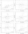
The relation between baseline EtCO2 (x‐axis) and the hemodynamic parameters (the CBF, the ASL CVR, the M value, the blood oxygenation level‐dependent CVR (ΔBOLD), the OEF and the CMRO2 is investigated. The relation between EtCO2 and CBF was significant (R 2 = 0.215, P < 0.01), same for the relation between EtCO2 and the CMRO2 (R 2 = 0.163, P < 0.01). There was no relation between EtCO2 and ASL CVR, M value, BOLD CVR or OEF (R 2 = 0.004, R 2 = 0.033, R 2 = 0.065, and R 2 = 0.015, respectively).
Figure 6
Figure 6
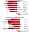
Results of the linear regression analysis. The graphs demonstrate the mean (95% confidence interval) difference in cerebral blood flow (CBF, in ml/100 g/min) and cerebral metabolic rate of oxygen (CMRO2, in µmol/100 g/min) between the young and old. The effect of EtCO2 as a confounder is given in light red, the remaining effect of the subgroup is shown in dark red. There remains a significant difference in the CMRO2 of the young versus elderly subjects measured in the frontal cortex, temporal cortex and the deep gray matter when EtCO2 is taken as a covariate. There is a trend toward a significant difference in whole brain gray matter CMRO2 (Wb GM, P = 0.063).
Figure 7
Figure 7
Scatter plot demonstrating the relation between the ASL CVR, defined as the percent change in CBF per 10 mmHg change in EtCO2, and the change in CBF per 1 mmHg change in EtCO2 (P < 0.05).

Similar articles

Cited by

References

    1. Aanerud J, Borghammer P, Chakravarty MM, Vang K, Rodell AB, Jonsdottir KY, Moller A, Ashkanian M, Vafaee MS, Iversen P, Johanssen P, Gjedde A (2012): Brain energy metabolism and blood flow differences in healthy aging. J Cereb Blood Flow Metab 32:1177–1187. - PMC - PubMed
    1. Alsop DC, Detre JA, Golay X, Gunther M, Hendrikse J, Hernandez‐Garcia L, Lu H, MacIntosh BJ, Parkes LM, Smits M, van Osch MJ, Wang DJ, Wong EC Zaharchuk G: Recommended implementation of arterial spin‐labeled perfusion MRI for clinical applications: A consensus of the ISMRM perfusion study group and the European consortium for ASL in dementia. Magn Reson Med (in press). doi: 10.1002/mrm.25197 - DOI - PMC - PubMed
    1. Anderson JM, Hubbard BM, Coghill GR, Slidders W (1983): The effect of advanced old age on the neurone content of the cerebral cortex. Observations with an automatic image analyser point counting method. J Neurol Sci 58:235–246. - PubMed
    1. Andersson JLR, Jenkinson M, Smith S (2007): Non‐Linear Registration, Aka Spatial Normalisation. FMRIB Technical Report TR07JA2. Available at: http://www.fmrib.ox.ac.uk/analysis/techrep
    1. Aslan S, Xu F, Wang PL, Uh J, Yezhuvath US, van Osch M, Lu H (2010): Estimation of labeling efficiency in pseudocontinuous arterial spin labeling. Magn Reson Med 63:765–771. doi:10.1002/mrm.25197. - DOI - PMC - PubMed

Publication types

MeSH terms

LinkOut - more resources