

Effects of lubricant and autologous bone marrow stromal cell augmentation on immobilized flexor tendon repairs
- PMID: 26177854
- PMCID: PMC5166703
- DOI: 10.1002/jor.22980
Effects of lubricant and autologous bone marrow stromal cell augmentation on immobilized flexor tendon repairs
Abstract
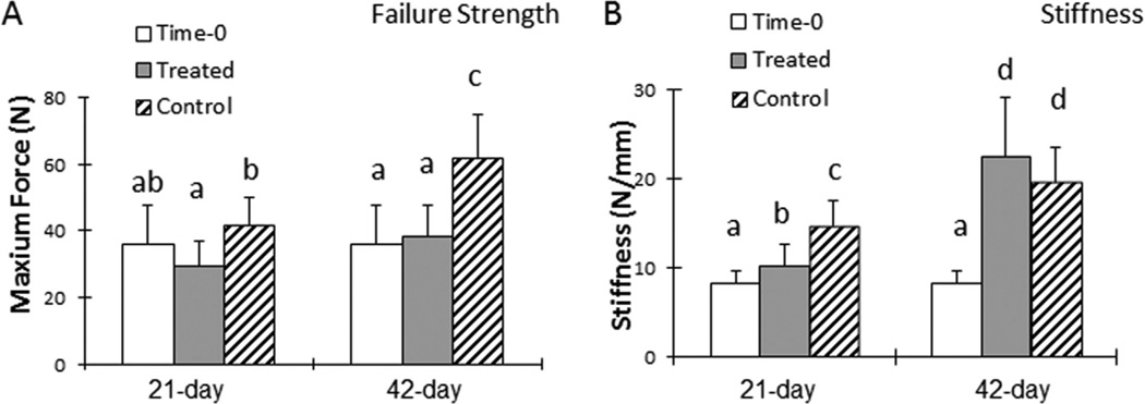
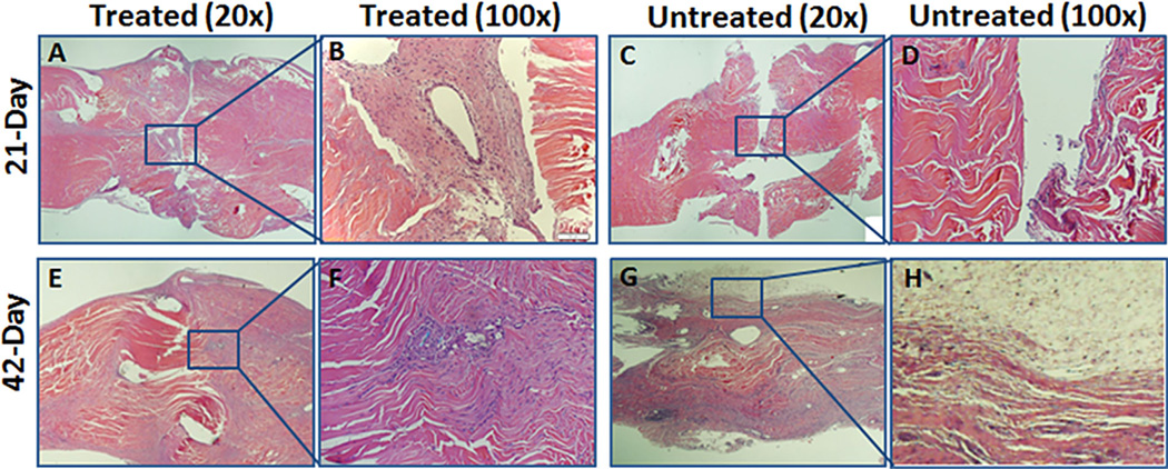
The purpose of the study was to test a novel treatment that carbodiimide-derivatized-hyaluronic acid-lubricin (cd-HA-lubricin) combined cell-based therapy in an immobilized flexor tendon repair in a canine model. Seventy-eight flexor tendons from 39 dogs were transected. One tendon was treated with cd-HA-lubricin plus an interpositional graft of 8 × 10(5) BMSCs and GDF-5. The other tendon was repaired without treatment. After 21 day of immobilization, 19 dogs were sacrificed; the remaining 20 dogs underwent a 21-day rehabilitation protocol before euthanasia. The work of flexion, tendon gliding resistance, and adhesion score in treated tendons were significantly less than the untreated tendons (p < 0.05). The failure strength of the untreated tendons was higher than the treated tendons at 21 and 42 days (p < 0.05). However, there is no significant difference in stiffness between two groups at day 42. Histologic analysis of treated tendons showed a smooth surface and viable transplanted cells 42 days after the repair, whereas untreated tendons showed severe adhesion formation around the repair site. The combination of lubricant and cell treatment resulted in significantly improved digit function, reduced adhesion formation. This novel treatment can address the unmet needs of patients who are unable to commence an early mobilization protocol after flexor tendon repair.
Keywords: bone marrow stromal cells; flexor tendon; hyaluronic acid; immobilization; lubricin; tendon repair.
© 2015 Orthopaedic Research Society. Published by Wiley Periodicals, Inc.
Figures


Similar articles
-
Resurfacing with chemically modified hyaluronic acid and lubricin for flexor tendon reconstruction.J Orthop Res. 2013 Jun;31(6):969-75. doi: 10.1002/jor.22305. Epub 2013 Jan 17. J Orthop Res. 2013. PMID: 23335124 Free PMC article.
-
Lubricin surface modification improves extrasynovial tendon gliding in a canine model in vitro.J Bone Joint Surg Am. 2008 Jan;90(1):129-35. doi: 10.2106/JBJS.G.00045. J Bone Joint Surg Am. 2008. PMID: 18171967
-
Lubricin surface modification improves tendon gliding after tendon repair in a canine model in vitro.J Orthop Res. 2009 Feb;27(2):257-63. doi: 10.1002/jor.20731. J Orthop Res. 2009. PMID: 18683890 Free PMC article.
-
Advances in the healing of flexor tendon injuries.Wound Repair Regen. 2014 May;22 Suppl 1:25-9. doi: 10.1111/wrr.12161. Wound Repair Regen. 2014. PMID: 24813361 Review.
-
[Graft reconstruction of flexor tendons].Chir Main. 2014 Dec;33 Suppl:S58-71. doi: 10.1016/j.main.2014.05.007. Epub 2014 Jul 8. Chir Main. 2014. PMID: 25026901 Review. French.
Cited by
-
Characterization of scar tissue biomechanics during adult murine flexor tendon healing.J Mech Behav Biomed Mater. 2022 Jun;130:105192. doi: 10.1016/j.jmbbm.2022.105192. Epub 2022 Mar 23. J Mech Behav Biomed Mater. 2022. PMID: 35339739 Free PMC article.
-
The revitalisation of flexor tendon allografts with bone marrow stromal cells and mechanical stimulation: An ex vivo model revitalising flexor tendon allografts.Bone Joint Res. 2017 Mar;6(3):179-185. doi: 10.1302/2046-3758.63.BJR-2016-0207.R1. Bone Joint Res. 2017. PMID: 28360084 Free PMC article.
-
Effect of connective tissue growth factor delivered via porous sutures on the proliferative stage of intrasynovial tendon repair.J Orthop Res. 2018 Jul;36(7):2052-2063. doi: 10.1002/jor.23842. Epub 2018 Feb 1. J Orthop Res. 2018. PMID: 29266404 Free PMC article.
-
Polydopamine Nanoparticles-Based Photothermal Effect Against Adhesion Formation in a Rat Model of Achilles Tendon Laceration Repair.Int J Nanomedicine. 2023 Apr 4;18:1765-1776. doi: 10.2147/IJN.S393454. eCollection 2023. Int J Nanomedicine. 2023. PMID: 37038441 Free PMC article.
-
Evaluating the Effectiveness of Commercially Available Antiadhesion Tendon Protector Sheets in Tendon Repair Surgery Versus Tendon Repair Surgery Alone: A Preclinical Model Study.J Hand Surg Am. 2025 Aug;50(8):1009.e1-1009.e10. doi: 10.1016/j.jhsa.2024.08.002. Epub 2024 Sep 21. J Hand Surg Am. 2025. PMID: 39306773
References
-
- Chesney A, Chauhan A, Kattan A, et al. Systematic review of flexor tendon rehabilitation protocols in zone II of the hand. Plastic Reconstr Surg. 2011;127:1583–1592. - PubMed
-
- Vucekovich K, Gallardo G, Fiala K. Rehabilitation after flexor tendon repair, reconstruction, and tenolysis. Hand Clin. 2005;21:257–265. - PubMed
-
- Strickland JW. Results of flexor tendon surgery in zone II. Hand Clin. 1985;1:167–179. - PubMed
-
- Foucher G, Lenoble E, Ben Youssef K, et al. A postoperative regime after digital flexor tenolysis. A series of 72 patients. J Hand Surg Eur. 1993;18:35–40. - PubMed
-
- Vahvanen V, Gripenberg L, Nuutinen P. Flexor tendon injury of the hand in children. A long-term follow-up study of 84 patients. Scand J Reconstr Surg. 1981;15:43–48. - PubMed
Publication types
MeSH terms
Substances
Grants and funding
LinkOut - more resources
Full Text Sources
Other Literature Sources
Medical
