

PHACE syndrome misdiagnosed as a port-wine stain
- PMID: 26177999
- PMCID: PMC4513526
- DOI: 10.1136/bcr-2015-209889
PHACE syndrome misdiagnosed as a port-wine stain
Erratum in
-
Correction.BMJ Case Rep. 2015 Aug 13;2015:bcr2015209889corr1. doi: 10.1136/bcr-2015-209889corr1. BMJ Case Rep. 2015. PMID: 26272956 Free PMC article. No abstract available.
Abstract
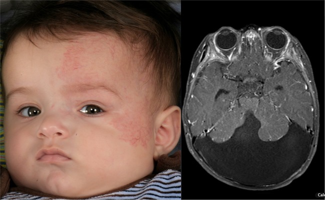
We present the case of a boy born with a large macular, segmental vascular anomaly over the left face, initially diagnosed as a capillary malformation (port-wine stain) by the postnatal paediatric team. The vascular anomaly in the face then grew rapidly during the first few weeks of life and started to occlude the left eye, causing parental concerns about the infant's vision. A dermatological opinion established that the lesion was a segmental infantile haemangioma (IH). This, in combination with the posterior fossa malformation previously detected on antenatal scanning and confirmed by an MRI postnatally, satisfied the criteria for Posterior fossa abnormalities, Haemangiomas, Arterial abnormalities, Cardiac abnormalities and Eye abnormalities (PHACE) syndrome: a rare cutaneous neurovascular syndrome. This case highlights the diagnostic challenge posed by early phenotypes of haemangiomas as well as the importance of correctly diagnosing PHACE syndrome.
2015 BMJ Publishing Group Ltd.
Figures

References
-
- Garzon M. Hemangiomas: update on classification, clinical presentation, and associated anomalies. Cutis 2000;66:325–8. - PubMed
Publication types
MeSH terms
Supplementary concepts
LinkOut - more resources
Full Text Sources
Other Literature Sources