Case Reports
. 2015 Jul 15:2015:bcr2015210717.
doi: 10.1136/bcr-2015-210717.

Congenital cystic eye with optic nerve

Affiliations
Case Reports

Congenital cystic eye with optic nerve

Lee Holland et al. BMJ Case Rep. .

Abstract

Congenital cystic eye (CCE) is a rare condition caused by failure of invagination of the optic vesicle resulting in a persistent cyst replacing the eye. An associated optic nerve attached to the cyst is a rarely reported phenomenon that has been sparsely described histologically, with no immunohistochemistry reported previously. The authors present a case of CCE with optic nerve tissue inserting into the cyst, and present the histological and immunohistochemical findings.


Figures

Figure 1
Figure 1
(A) Patient at 1 month of age with right congenital cystic eye (CCE) visible as a blue-tinged orbital swelling displacing the normal right lower lid upwards. The left eye was normal. (B) Well-circumscribed, heterogeneous, cystic mass in the right orbit, which is hypointense to cerebrospinal fluid on coronal T2-weighted MRI. (C) Axial CT image demonstrating a calcific area within the cyst. The lacrimal gland, orbital bones, extraocular muscle cone and optic chiasm had normal appearances. (D) An axial CT image at a different anatomical level showed a normal medial rectus muscle, lateral rectus muscle and optic nerve-like structure merging with the wall of the cyst. (E) Macroscopic appearance of CCE immediately after surgical removal. Arrow denotes area of insertion of optic nerve. (F) Aperio scan of a complete transverse section through the CCE (24 mm×18 mm), with the optic nerve attached posteriorly (arrow). An area of dystrophic calcification is seen within the cavity (arrowhead) corresponding to the radiological findings.
Figure 2
Figure 2
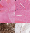
(A) Gliotic tissue within congenital cystic eye (H&E, ×200). (B) Glial fibrillary acidic protein (GFAP) highlighting glial tissue within congenital cystic eye (CCE) (×100). (C) Disorganised retinal tissue in congenital cystic eye (H&E, ×100). (D) Synaptophysin highlighting retinal cells within CCE (×200).
Figure 3
Figure 3
(A) Transverse section of optic nerve showing relationship to retinal vessels (H&E, ×40). (B) Transverse section of optic nerve showing central retinal vein (H&E, ×200). (C) Longitudinal section of optic nerve attaching to the glial tissue in the CCE (H&E, ×40). (D) Longitudinal section of optic nerve showing central retinal vessel (H&E, ×100). (E) Optic nerve immunohistochemistry for glial fibrillary acidic protein (GFAP) (×100). (F) Meningeal tissue adjacent to optic nerve positive for epithelial membrane antigen (EMA, ×100).

References

    1. Mann I. A case of congenital cystic eye. Trans Ophthalmol Soc Aust 1939;1:120–4.
    1. Harlan GC. Case of congenital cyst of the orbit with anophthalmos. Trans Am Ophthalmol Soc 1893;6:559–65. - PMC - PubMed
    1. Gangadhar JL, Indiradevi B, Prabhakaran VC. Congenital cystic eye with meningocele. J Pediatr Neurosci 2009;4:136–8. 10.4103/1817-1745.57334 - DOI - PMC - PubMed
    1. Gupta R, Seith A, Guglani B et al. . Congenital cystic eye: features on MRI. Br J Radiol 2007;80:e137–40. 10.1259/bjr/31817019 - DOI - PubMed
    1. Kavanagh MC, Tam D, Diehn JJ et al. . Detection of a congenital cystic eyeball by prenatal ultrasound in a newborn with Turner's syndrome. Br J Ophthalmol 2007;91:559–60. 10.1136/bjo.2006.096107 - DOI - PMC - PubMed

Publication types