. 2015 Sep 18;290(38):22955-69.
doi: 10.1074/jbc.M115.660183. Epub 2015 Jul 15.

Structural and Functional Characterization of a Lytic Polysaccharide Monooxygenase with Broad Substrate Specificity

Affiliations

Structural and Functional Characterization of a Lytic Polysaccharide Monooxygenase with Broad Substrate Specificity

Anna S Borisova et al. J Biol Chem. .

Abstract

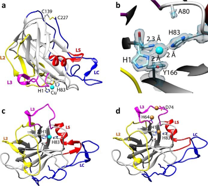
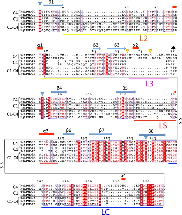
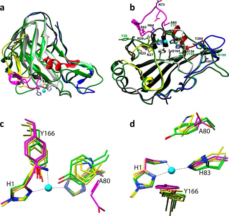
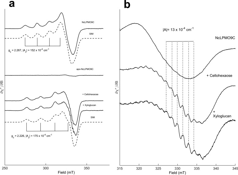
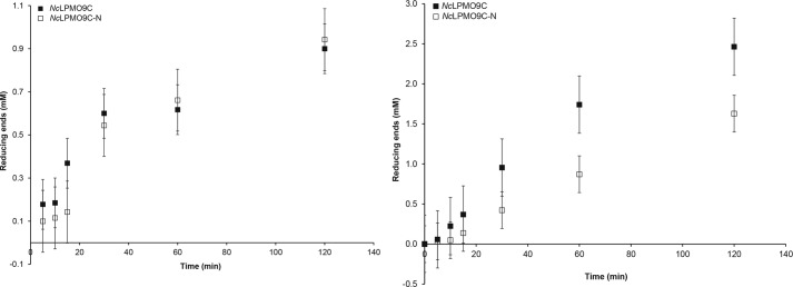
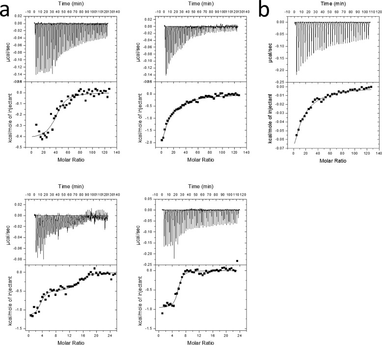
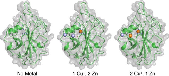
The recently discovered lytic polysaccharide monooxygenases (LPMOs) carry out oxidative cleavage of polysaccharides and are of major importance for efficient processing of biomass. NcLPMO9C from Neurospora crassa acts both on cellulose and on non-cellulose β-glucans, including cellodextrins and xyloglucan. The crystal structure of the catalytic domain of NcLPMO9C revealed an extended, highly polar substrate-binding surface well suited to interact with a variety of sugar substrates. The ability of NcLPMO9C to act on soluble substrates was exploited to study enzyme-substrate interactions. EPR studies demonstrated that the Cu(2+) center environment is altered upon substrate binding, whereas isothermal titration calorimetry studies revealed binding affinities in the low micromolar range for polymeric substrates that are due in part to the presence of a carbohydrate-binding module (CBM1). Importantly, the novel structure of NcLPMO9C enabled a comparative study, revealing that the oxidative regioselectivity of LPMO9s (C1, C4, or both) correlates with distinct structural features of the copper coordination sphere. In strictly C1-oxidizing LPMO9s, access to the solvent-facing axial coordination position is restricted by a conserved tyrosine residue, whereas access to this same position seems unrestricted in C4-oxidizing LPMO9s. LPMO9s known to produce a mixture of C1- and C4-oxidized products show an intermediate situation.

Keywords: biodegradation; bioenergy; copper monooxygenase; crystal structure; electron paramagnetic resonance (EPR); isothermal titration calorimetry (ITC).


Figures

FIGURE 1.
FIGURE 1.
Structural representations of NcLPMO9C-N. a, cartoon representation of the copper-bound structure; copper is depicted as a cyan sphere; b, close up of the copper-binding site with the electron density map around the active site in gray mesh (contoured at 1σ); c, overall structure of the copper-loaded protein rotated by 90° along the horizontal axis compared with the view in a; d, structure of the zinc-loaded protein with the three bound zinc ions depicted as brown spheres; the orientation is similar to that in c. Note the structural variation in the loop coordinating the third zinc ion (residues 70–76 in the L3 loop, colored in pink).
FIGURE 2.
FIGURE 2.
Structure-based sequence alignment of LPMO9s with known structures. The proteins included are as follows: NcLPMO9C-N (PDB code 4D7U), NcLPMO9D (PDB code 4EIR), TtLPMO9E (PDB code 3EII), PcLPMO9D (PDB code 4B5Q), TaLPMO9A (PDB code 3ZUD), NcLPMO9M (PDB code 4EIS), and HjLPMO9B (PDB code 2VTC). Fully conserved residues are shown in white on a red background. Blue frames indicate that more than 70% of the residues in the corresponding columns exhibit similar physico-chemical properties (indicated as red residues on a white background). Blue triangles indicate residues coordinating the active site metal, and yellow triangles indicate residues involved in binding of two additional zinc ions. The secondary structure assignment (β-strands indicated as blue arrows and α-helices as red cylinders) refers to NcLPMO9C-N and was determined with the program DSSP (56). The oxidative regio-specificity of the LPMOs, indicated on the left, was assigned based on experimental evidence (4, 5, 13, 57) or, for HjLPMO9B and TtLPMO9E, by inference from the sequence-based categorization (43, 45). The residue numbered 80, which affects the accessibility of the solvent-facing axial copper coordination site, as shown in Fig. 4, c and d, and discussed in detail in the text, is indicated by a black asterisk. The loop regions that contribute to shaping the substrate-binding surface, named L2, LC, L3, and LS (see text), are marked by horizontal bars below the sequence, with color coding as in Fig. 2. The figure was prepared with ESPript.
FIGURE 3.
FIGURE 3.
Structural comparison of LPMO9s. a, superposition of PcGH61D (green; PDB code 4B5Q) with NcLPMO9C-N (colored as in Fig. 2). b, comparison of protruding surface residues in NcLPMO9C (colored as in a) with residues at equivalent positions in C1-oxidizing PcLPMO9D (green). b is rotated 90° along the horizontal axis compared with a. c and d, superposition of the copper sites of seven LPMO9s with known structure; C1, C1/C4, and C4 oxidizers are colored green, yellow, and magenta, respectively. The orientation shown in c is similar to that in a, whereas the orientation in d resembles that of b. Residue numbers refer to NcLPMO9C, with the exception of green labels in b, which refer to PcLPMO9D. The copper in NcLPMO9C is shown as a cyan sphere in all panels.
FIGURE 4.
FIGURE 4.
NcLPMO9C EPR spectra. a, EPR spectra of 160 μm Cu2+-loaded NcLPMO9C in the absence (upper panel) or presence (lower panel) of the soluble substrates cellohexaose (20 mg/ml) or xyloglucan (15 mg/ml). Spectra were recorded at 77 K, 1 milliwatt of microwave power, and 10 gauss modulation amplitude. The simulated spectra (SIM) for each species are shown below the corresponding experimental spectra (simulation parameters for the cellohexaose and xyloglucan spectra were identical). b, effect of substrate on super hyperfine splitting in the high field region. The splitting constants (|A| ∼13 × 10−4 cm) are consistent with coupling to nearby nitrogen nuclei. Spectra were recorded at 77 K, 1 milliwatt of microwave power, and 2 gauss modulation amplitude.
FIGURE 5.
FIGURE 5.
Comparison of substrate degradation rates. Degradation of 5 mg/ml PASC (left) or tamarind xyloglucan (right) by 4 μm NcLPMO9C with or without (−N) the CBM domain at 50 °C was monitored by measuring the formation of reducing ends. In the absence of a reductant, the enzyme reactions did not yield reducing ends (not shown).
FIGURE 6.
FIGURE 6.
Thermograms. a, upper panels, binding isotherms with theoretical fits (lower panels) obtained for the titration of NcLPMO9C (left) and NcLPMO9C-N (right) into 0.9 μm xyloglucan (top) and 0.146 mg/ml PASC (bottom). The concentration of PASC was set to be 4.5 μm based on an estimated degree of polymerization of 200 (38). All experiments were carried out at pH 5.5. The temperature was 25 °C for PASC and 10 °C for xyloglucan; b, upper panel, binding isotherms with theoretical fits (lower panel) obtained for the binding of 11 mm Glc6 to 30 μm NcLPMO9C-Cu2+ at t = 25 °C.
FIGURE 7.
FIGURE 7.
Illustration of the three different bound metal scenarios considered in the computational docking study of NcLPMO9C-N.

References

    1. Horn S. J., Vaaje-Kolstad G., Westereng B., Eijsink V. G. (2012) Novel enzymes for the degradation of cellulose. Biotechnol. Biofuels 5, 45. - PMC - PubMed
    1. Forsberg Z., Vaaje-Kolstad G., Westereng B., Bunæs A. C., Stenstrøm Y., MacKenzie A., Sørlie M., Horn S. J., Eijsink V. G. (2011) Cleavage of cellulose by a CBM33 protein. Protein Sci. 20, 1479–1483 - PMC - PubMed
    1. Harris P. V., Welner D., McFarland K. C., Re E., Navarro Poulsen J. C., Brown K., Salbo R., Ding H., Vlasenko E., Merino S., Xu F., Cherry J., Larsen S., Lo Leggio L. (2010) Stimulation of lignocellulosic biomass hydrolysis by proteins of glycoside hydrolase family 61: structure and function of a large, enigmatic family. Biochemistry 49, 3305–3316 - PubMed
    1. Phillips C. M., Beeson W. T., Cate J. H., Marletta M. A. (2011) Cellobiose dehydrogenase and a copper-dependent polysaccharide monooxygenase potentiate cellulose degradation by Neurospora crassa. ACS Chem. Biol. 6, 1399–1406 - PubMed
    1. Quinlan R. J., Sweeney M. D., Lo Leggio L., Otten H., Poulsen J. C., Johansen K. S., Krogh K. B., Jørgensen C. I., Tovborg M., Anthonsen A., Tryfona T., Walter C. P., Dupree P., Xu F., Davies G. J., Walton P. H. (2011) Insights into the oxidative degradation of cellulose by a copper metalloenzyme that exploits biomass components. Proc. Natl. Acad. Sci. U.S.A. 108, 15079–15084 - PMC - PubMed

Publication types

MeSH terms