

Cutaneous Intravascular NK/T-cell lymphoma mimic panniculitis clinically, case report and literature brief review
- PMID: 26178620
- PMCID: PMC4504160
- DOI: 10.1186/s13000-015-0330-0
Cutaneous Intravascular NK/T-cell lymphoma mimic panniculitis clinically, case report and literature brief review
Abstract
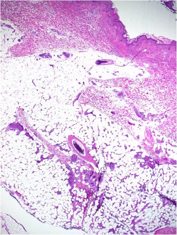
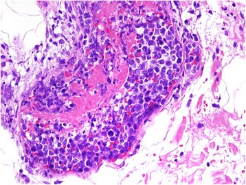
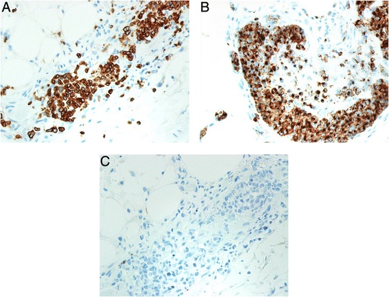
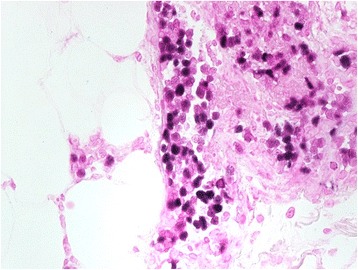
Intravascular large cell lymphoma is a rare subtype of extranodal large cell lymphoma characterized by the presence of neoplastic cells within the lumina of small vessels. Most cases of intravascular large cell lymphoma have a B-cell phenotype. To date, 12 cases of intravascular natural killer (NK/)/T-cell lymphoma (IVNKL) have been reported. Our case is A 47-year-old female presented with erythematous patches and plaques on the lower extremities mimicking panniculitis clinically. A skin biopsy revealed intravascular lymphoma (IVL) with a NK/T cell phenotype (positive for CD3, and granzyme B and negative for CD20, CD4, CD8, CD5). The lymphoma cells were also positive for Epstein-Barr virus by Epstein-Barr virus-encoded RNA in situ hybridization test. Because this type of lymphoma is extremely rare, our case is documented and compared with the previously reported cases.
Figures
References
-
- Pfleger L, Tappeiner J. On the recognition of systematized endotheliomatosis of the cutaneous blood vessels (reticuloendotheliosis?) Hautarzt. 1959;10:359–363. - PubMed
-
- Gatter KC, Warnke RA. Intravascular large B-cell lymphoma: World Health Organization Classification of Tumors. Pathology and Genetics of Tumours of Haematopoietic and Lymphoid Tissues. Lyon, France: IARC Press; 2001. pp. 177–178.
Publication types
MeSH terms
Substances
LinkOut - more resources
Full Text Sources
Other Literature Sources
Medical
Research Materials
