

Ileal mucosa-associated lymphoid tissue lymphoma presenting with small bowel obstruction: a case report
- PMID: 26178711
- PMCID: PMC4504228
- DOI: 10.1186/s13000-015-0353-6
Ileal mucosa-associated lymphoid tissue lymphoma presenting with small bowel obstruction: a case report
Abstract
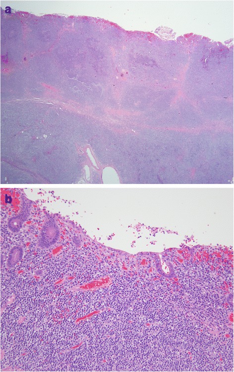
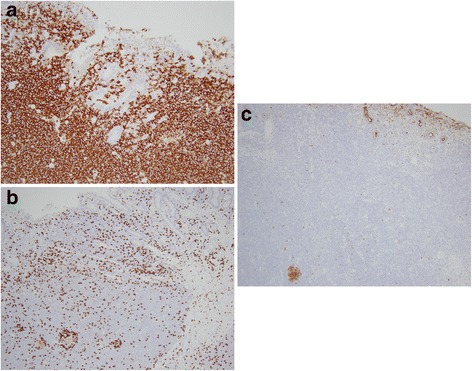
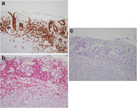
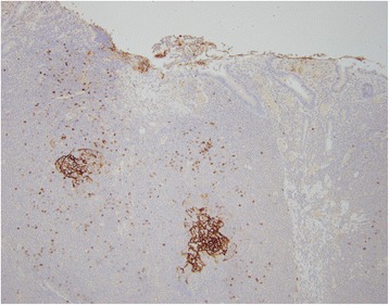
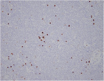
Extranodal marginal zone lymphoma of mucosa-associated lymphoid tissue (MALT Lymphoma) of the gastrointestinal tract commonly involves the stomach in the setting of concurrent Helicobacter pylori (H. pylori) infection. Primary ileal MALT lymphoma is rare, and has not been associated with a specific infectious disease. We report a case of a 58-year-old man who presented to the emergency department with constipation and abdominal distension, and signs of an obstructing mass on computed tomography scan. A small bowel resection was performed which revealed an 8 cm saccular dilatation with thickened bowel wall and subjacent thickened tan-yellow tissue extending into the mesentery. Histologically, there was a diffuse lymphoid infiltrate consisting of small atypical cells with monocytoid features. These cells were CD20-positive B-lymphocytes that co-expressed BCL-2 and were negative for CD5, CD10, CD43, and cyclin D1 on immunohistochemical studies. Kappa-restricted plasma cells were also identified by in situ hybridization. The overall proliferation index was low with Ki-67 immunoreactivity in approximately 10 % of cells. No areas suspicious for large cell or high grade transformation were identified. The pathologic findings were diagnostic of an extranodal marginal zone lymphoma involving the ileum, with early involvement of mesenteric lymph nodes. Small hypermetabolic right mesenteric and bilateral hilar lymph nodes were identified by imaging. The bone marrow biopsy showed no evidence of involvement by lymphoma. The patient was clinically considered advanced stage and opted for therapy with rituximab infusions. After six months of therapy, follow-up radiologic studies demonstrated significant decrease in size of the mesenteric lymph nodes.
Figures

Similar articles
-
Extranodal marginal zone lymphoma of mucosa-associated lymphoid tissue (MALT lymphoma) of the ileum in a 35-year-old Japanese woman.Int J Clin Exp Pathol. 2013 Apr 15;6(5):951-6. Print 2013. Int J Clin Exp Pathol. 2013. PMID: 23638229 Free PMC article.
-
Hodgkin's disease and an extranodal marginal zone B-cell lymphoma in the small intestine: an unusual composite lymphoma.Mod Pathol. 1996 Oct;9(10):1020-6. Mod Pathol. 1996. PMID: 8902841
-
[Ileus of the small intestine in intestinal marginal-zone B-cell lymphoma of mucoid-associated lymphoid tissue (MALT)].Dtsch Med Wochenschr. 2001 Dec 7;126(49):1391-5. doi: 10.1055/s-2001-18881. Dtsch Med Wochenschr. 2001. PMID: 11740631 German.
-
Synchronous colonic adenoma and intestinal marginal zone B-cell lymphoma associated with Crohn's disease: a case report and literature review.BMC Cancer. 2019 Oct 17;19(1):966. doi: 10.1186/s12885-019-6224-x. BMC Cancer. 2019. PMID: 31623635 Free PMC article. Review.
-
Isolated gastric amyloidoma in the setting of marginal zone MALT lymphoma: case report and review of the literature.Conn Med. 2014 May;78(5):277-80. Conn Med. 2014. PMID: 24974561 Review.
Cited by
-
Mucosa-Associated Lymphoid Tissue (MALT) Lymphoma in the Gastrointestinal Tract in the Modern Era.Cancers (Basel). 2022 Jan 17;14(2):446. doi: 10.3390/cancers14020446. Cancers (Basel). 2022. PMID: 35053607 Free PMC article. Review.
-
Challenges in the diagnosis of marginal zone lymphoma with symptoms of small intestinal disease: a case report and scoping review of the literature.J Gastrointest Oncol. 2022 Oct;13(5):2583-2607. doi: 10.21037/jgo-22-74. J Gastrointest Oncol. 2022. PMID: 36388684 Free PMC article.
-
Small Bowel Obstruction Secondary to Ileal Mucosa-Associated Lymphoid Tissue Lymphoma.J Gastrointest Cancer. 2018 Jun;49(2):207-210. doi: 10.1007/s12029-016-9882-9. J Gastrointest Cancer. 2018. PMID: 27726063 No abstract available.
-
Rare presentation of ileal mucosa-associated lymphoid tissue lymphoma: a case of obstructive syndrome in a Moroccan patient.J Surg Case Rep. 2025 Jul 14;2025(7):rjaf534. doi: 10.1093/jscr/rjaf534. eCollection 2025 Jul. J Surg Case Rep. 2025. PMID: 40661721 Free PMC article.
-
Whole-Exome Sequencing Reveals New Potential Mutations Genes for Primary Mucosa-Associated Lymphoid Tissue Lymphoma Arising From the Kidney.Front Oncol. 2021 Jan 8;10:609839. doi: 10.3389/fonc.2020.609839. eCollection 2020. Front Oncol. 2021. PMID: 33585230 Free PMC article.
References
Publication types
MeSH terms
Substances
LinkOut - more resources
Full Text Sources
Other Literature Sources
Medical
Research Materials
