Developmental Programming: Does Prenatal Steroid Excess Disrupt the Ovarian VEGF System in Sheep?
- PMID: 26178718
- PMCID: PMC4710184
- DOI: 10.1095/biolreprod.115.131607
Developmental Programming: Does Prenatal Steroid Excess Disrupt the Ovarian VEGF System in Sheep?
Abstract
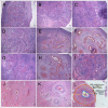
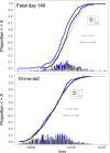
Prenatal testosterone (T), but not dihydrotestosterone (DHT), excess disrupts ovarian cyclicity and increases follicular recruitment and persistence. We hypothesized that the disruption in the vascular endothelial growth factor (VEGF) system contributes to the enhancement of follicular recruitment and persistence in prenatal T-treated sheep. The impact of T/DHT treatments from Days 30 to 90 of gestation on VEGFA, VEGFB, and their receptor (VEGFR-1 [FLT1], VEGFR-2 [KDR], and VEGFR-3 [FLT4]) protein expression was examined by immunohistochemistry on Fetal Days 90 and 140, 22 wk, 10 mo (postpubertal), and 21 mo (adult) of age. Arterial morphometry was performed in Fetal Day 140 and postpubertal ovaries. VEGFA and VEGFB expression were found in granulosa cells at all stages of follicular development with increased expression in antral follicles. VEGFA was present in theca interna, while VEGFB was present in theca interna/externa and stromal cells. All three receptors were expressed in the granulosa, theca, and stromal cells during all stages of follicular development. VEGFR-3 increased with follicular differentiation with the highest level seen in the granulosa cells of antral follicles. None of the members of the VEGF family or their receptor expression were altered by age or prenatal T/DHT treatments. At Fetal Day 140, area, wall thickness, and wall area of arteries from the ovarian hilum were larger in prenatal T- and DHT-treated females, suggestive of early androgenic programming of arterial differentiation. This may facilitate increased delivery of endocrine factors and thus indirectly contribute to the development of the multifollicular phenotype.
Keywords: PCOS; VEGF; ovary; sheep; testosterone.
© 2015 by the Society for the Study of Reproduction, Inc.
Figures


Similar articles
-
Developmental Programming: Prenatal Testosterone Excess on Ovarian SF1/DAX1/FOXO3.Reprod Sci. 2020 Jan;27(1):342-354. doi: 10.1007/s43032-019-00029-0. Epub 2020 Jan 1. Reprod Sci. 2020. PMID: 32046386 Free PMC article.
-
Developmental programming: prenatal steroid excess disrupts key members of intraovarian steroidogenic pathway in sheep.Endocrinology. 2014 Sep;155(9):3649-60. doi: 10.1210/en.2014-1266. Epub 2014 Jul 25. Endocrinology. 2014. PMID: 25061847 Free PMC article.
-
Developmental programming: differential effects of prenatal testosterone and dihydrotestosterone on follicular recruitment, depletion of follicular reserve, and ovarian morphology in sheep.Biol Reprod. 2009 Apr;80(4):726-36. doi: 10.1095/biolreprod.108.072801. Epub 2008 Dec 17. Biol Reprod. 2009. PMID: 19092114 Free PMC article.
-
Steroidogenic versus Metabolic Programming of Reproductive Neuroendocrine, Ovarian and Metabolic Dysfunctions.Neuroendocrinology. 2015;102(3):226-37. doi: 10.1159/000381830. Epub 2015 Apr 1. Neuroendocrinology. 2015. PMID: 25832114 Free PMC article. Review.
-
Intra-follicular activin availability is altered in prenatally-androgenized lambs.Mol Cell Endocrinol. 2001 Dec 20;185(1-2):51-9. doi: 10.1016/s0303-7207(01)00632-3. Mol Cell Endocrinol. 2001. PMID: 11738794 Review.
Cited by
-
High-fat diet-induced dysregulation of ovarian gene expression is restored with chronic omega-3 fatty acid supplementation.Mol Cell Endocrinol. 2020 Jan 1;499:110615. doi: 10.1016/j.mce.2019.110615. Epub 2019 Oct 16. Mol Cell Endocrinol. 2020. PMID: 31628964 Free PMC article.
-
Developmental programming: prenatal testosterone-induced epigenetic modulation and its effect on gene expression in sheep ovary†.Biol Reprod. 2020 Apr 24;102(5):1045-1054. doi: 10.1093/biolre/ioaa007. Biol Reprod. 2020. PMID: 31930385 Free PMC article.
-
Animal Models to Understand the Etiology and Pathophysiology of Polycystic Ovary Syndrome.Endocr Rev. 2020 Jul 1;41(4):bnaa010. doi: 10.1210/endrev/bnaa010. Endocr Rev. 2020. PMID: 32310267 Free PMC article. Review.
-
Effects of Elevated Maternal Adiposity on Offspring Reproductive Health: A Perspective From Epidemiologic Studies.J Endocr Soc. 2022 Oct 27;7(1):bvac163. doi: 10.1210/jendso/bvac163. eCollection 2022 Nov 17. J Endocr Soc. 2022. PMID: 36438545 Free PMC article. Review.
-
Role of vascular endothelial growth factor B in nonalcoholic fatty liver disease and its potential value.World J Hepatol. 2023 Jun 27;15(6):786-796. doi: 10.4254/wjh.v15.i6.786. World J Hepatol. 2023. PMID: 37397934 Free PMC article. Review.
References
-
- Zeleznik AJ, Schuler HM, Reichert LE., Jr. Gonadotropin-binding sites in the rhesus monkey ovary: role of the vasculature in the selective distribution of human chorionic gonadotropin to the preovulatory follicle. Endocrinology. 1981;109:356–362. - PubMed
-
- Greenwald GS. Temporal and topographic changes in DNA synthesis after induced follicular atresia. Biol Reprod. 1989;41:175–181. - PubMed
-
- Ferrara N, Chen H, Davis-Smyth T, Gerber H-P, Nguyen T-N, Peers D, Chisholm V, Hillan KJ, Schwall RH. Vascular endothelial growth factor is essential for corpus luteum angiogenesis. Nat Med. 1998;4:336–340. - PubMed
-
- Fraser HM, Dickson SE, Lunn SF, Wulff C, Morris KD, Carroll VA, Bicknell R. Suppression of luteal angiogenesis in the primate after neutralization of vascular endothelial growth factor. Endocrinology. 2000;141:995–1000. - PubMed
-
- Wulff C, Wiegand SJ, Saunders PT, Scobie GA, Fraser HM. Angiogenesis during follicular development in the primate and its inhibition by treatment with truncated Flt-1-Fc (vascular endothelial growth factor Trap(A40)) Endocrinology. 2001;142:3244–3254. - PubMed
Publication types
MeSH terms
Substances
Grants and funding
LinkOut - more resources
Full Text Sources
Other Literature Sources
Medical
Miscellaneous

