

Pulmonary epithelioid inflammatory myofibroblastic sarcoma with multiple bone metastases: case report and review of literature
- PMID: 26178751
- PMCID: PMC4504132
- DOI: 10.1186/s13000-015-0358-1
Pulmonary epithelioid inflammatory myofibroblastic sarcoma with multiple bone metastases: case report and review of literature
Abstract
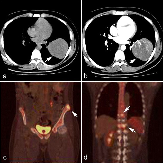
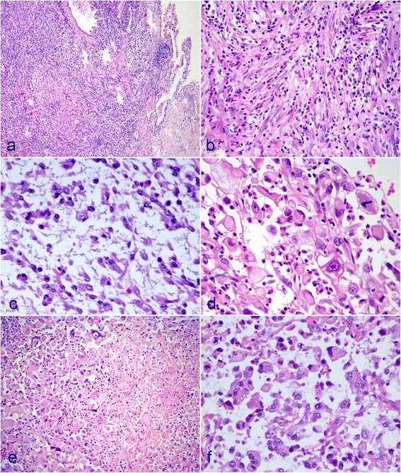
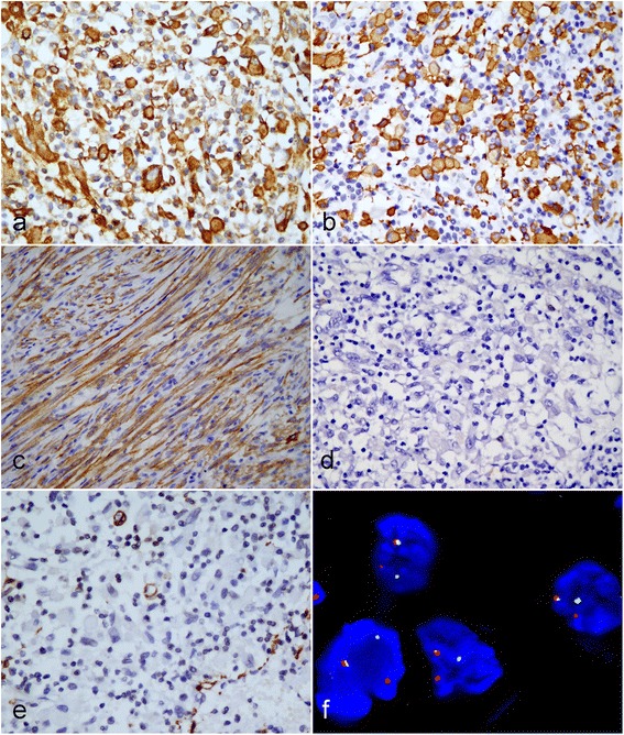
Epithelioid inflammatory myofibroblastic sarcoma (EIMS) is a rare variant of inflammatory myofibroblastic tumor with distinctive morphological features and malignant clinical behavior. Only a few such cases have been described in the literature. We report here a case of unusual pulmonary EIMS with multiple bone metastases. A 21-year-old Chinese male patient presented with complaints of general fatigue and rapid weight loss, and a huge tumor arising in the left lower lobe of lung was detected by chest computed tomography. The mass of lung was totally resected. Microscopically, the tumor cells were rounded and epithelioid in shape. Myxoid stroma and inflammatory infiltration was also present. The tumor cells were immunopositive to anaplastic lymphoma kinase (ALK) in smooth cytoplasmic pattern. Fluorescence in situ hybridization (FISH) assay revealed the presence of rearrangement of ALK gene. Three months after lung surgery, there were multiple bone metastases and intraspinal mass found by positron emission tomography. The second surgical treatment was performed to remove the intraspinal lesion. The histological and immunohistochemical features of intraspinal mass were similar to those of pulmonary tumor. The diagnosis of pulmonary EIMS with multiple bone metastases was made. To the best of our knowledge, it may be the first case of an EIMS arising in lung. Awareness of EIMS in respiratory tract and its distinctive features is important for pathologists to avoid a diagnostic pitfall caused by histologic similarities to other ALK-positive tumors. ALK inhibitor is a promising treatment for this aggressive tumor regardless of its potential acquired resistance.
Figures
References
-
- Coffin CM, Fletcher JA. Inflammatory myofibroblastic tumor. In: Fletcher CDM, Bridge JA, Hogendoorn PCW, Mertens F, editors. World Health Organization classification of tumors of soft tissue and bone. Lyon: IARC Press; 2013. pp. 83–4.
-
- Mariño-Enríquez A, Wang WL, Roy A, Lopez-Terrada D, Lazar AJ, Fletcher CD, et al. Epithelioid inflammatory myofibroblastic sarcoma: An aggressive intra-abdominal variant of inflammatory myofibroblastic tumor with nuclear membrane or perinuclear ALK. Am J Surg Pathol. 2011;35:135–44. doi: 10.1097/PAS.0b013e318200cfd5. - DOI - PubMed
-
- Suzuki R, Seto M, Nakamura S, Nakagawa A, Hara K, Takeuchi K. Sarcomatoid variant of anaplastic large cell lymphoma with cytoplasmic ALK and alpha-smooth muscle actin expression: a mimic of inflammatory myofibroblastic tumor. Am J Pathol. 2001;159:383–4. doi: 10.1016/S0002-9440(10)61706-3. - DOI - PMC - PubMed
Publication types
MeSH terms
Substances
LinkOut - more resources
Full Text Sources
Other Literature Sources
Medical
