. 2016 Jan;77(12):1611-6.
doi: 10.1292/jvms.15-0121. Epub 2015 Jul 16.

The effect of glycosylation on cytotoxicity of Ibaraki virus nonstructural protein NS3

Affiliations

The effect of glycosylation on cytotoxicity of Ibaraki virus nonstructural protein NS3

Maho Urata et al. J Vet Med Sci. 2016 Jan.

Abstract

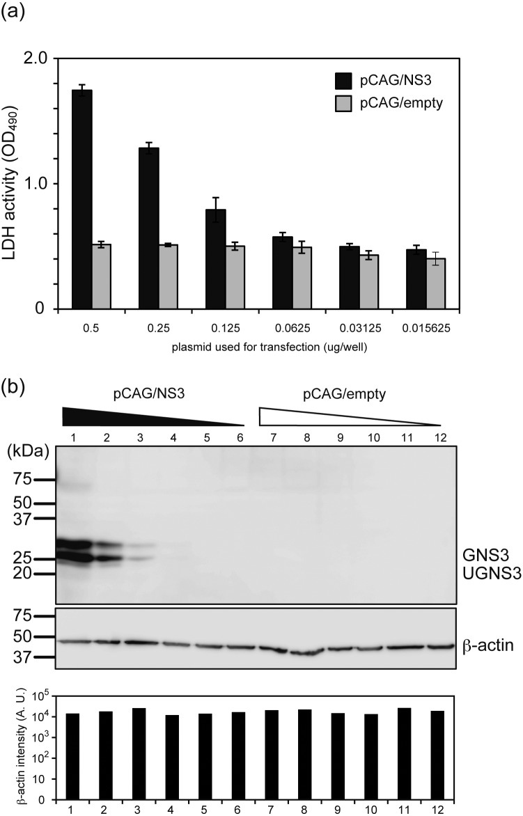
The cytotoxicity of Ibaraki virus nonstructural protein NS3 was confirmed, and the contribution of glycosylation to this activity was examined by using glycosylation mutants of NS3 generated by site-directed mutagenesis. The expression of NS3 resulted in leakage of lactate dehydrogenase to the culture supernatant, suggesting the cytotoxicity of this protein. The lack of glycosylation impaired the transport of NS3 to the plasma membrane and resulted in reduced cytotoxicity. Combined with the previous observation that NS3 glycosylation was specifically observed in mammalian cells (Urata et al., Virus Research 2014), it was suggested that the alteration of NS3 cytotoxicity through modulating glycosylation is one of the strategies to achieve host specific pathogenisity of Ibaraki virus between mammals and vector arthropods.


Figures

Fig. 1.
Fig. 1.
The alignment of NS3 amino acid sequence between IBAV and BTV10. The deduced amino acid sequence of IBAV NS3 was aligned with BTV NS3 by the ClastalW2 program. Putative N-linked glycosylation sites are underlined, predicted transmembrane domains are indicated by bold, and viral late domains are surrounded by square.
Fig. 2.
Fig. 2.
Production of NS3 glycosylation mutants. (a) Schematic illustration of NS3 glycosylation mutants generated in this study. Gray area represents the transmembrane domain. (b) Western blot analysis of 293T cells expressing wt-NS3, NS3/N59Q, NS3/N145Q and NS3/N59,145Q. NS3 was detected by western blot analysis using mouse anti-NS3 sera with both glycosylated (GNS3) and unglycosylated (UGNS3) proteins indicated.
Fig. 3.
Fig. 3.
IBAV NS3 possesses cytotoxicity on mammalian cells. (a) LDH activity in the culture supernatant of 293T cells transfected with various amounts of wt-NS3 expression plasmid (black bar) or control plasmid pCAG/empty (gray bar). The result is the representative of three independent experiments. (b) Western blot analysis of cells used for LDH assay with anti-NS3 and anti-β-actin antibodies. The bottom graph shows the intensity of β-actin protein band quantified by ImageJ software.
Fig. 4.
Fig. 4.
NS3 glycosylation mutants showed lower cytotoxicity in mammalian cells compared to wt NS3. (a) LDH activity in the culture supernatant of 293T cells transfected with expression plasmids for wt-NS3 and various glycosylation mutants. The result is the representative of three independent experiments. n=6 for each experiment. (*P<0.001) (b) Western blot analysis of cells used for LDH assay with anti-NS3 and anti-β-actin antibodies. The middle graph shows the intensity of total NS3 (GNS3 plus UGNS3) quantified by ImageJ software.
Fig. 5.
Fig. 5.
Visualization of NS3 in 293T cells expressing wt-NS3 and NS3 glycosylation mutants by immunofluorescence assay. (a) pCA7/NS3 and (b) pCA7/NS3N59,145Q were transfected to 293T cells. NS3 was visualized by IFA with protein specific antisera. The longitudinal and cross-section images were also created. NS3, the plasma membrane and the nucleus are shown in green, red and blue, respectively. Co-localization of NS3 and the plasma membrane is shown in yellow.

References

    1. Anthony S. J., Maan N., Maan S., Sutton G., Attoui H., Mertens P. P.2009. Genetic and phylogenetic analysis of the non-structural proteins NS1, NS2 and NS3 of epizootic haemorrhagic disease virus (EHDV). Virus Res. 145: 211–219. doi: 10.1016/j.virusres.2009.07.019 - DOI - PubMed
    1. Bansal O. B., Stokes A., Bansal A., Bishop D., Roy P.1998. Membrane organization of bluetongue virus nonstructural glycoprotein NS3. J. Virol. 72: 3362–3369. - PMC - PubMed
    1. Beaton A. R., Rodriguez J., Reddy Y. K., Roy P.2002. The membrane trafficking protein calpactin forms a complex with bluetongue virus protein NS3 and mediates virus release. Proc. Natl. Acad. Sci. U.S.A. 99: 13154–13159. doi: 10.1073/pnas.192432299 - DOI - PMC - PubMed
    1. Celma C. C., Roy P.2011. Interaction of calpactin light chain (S100A10/p11) and a viral NS protein is essential for intracellular trafficking of nonenveloped bluetongue virus. J. Virol. 85: 4783–4791. doi: 10.1128/JVI.02352-10 - DOI - PMC - PubMed
    1. Celma C. C., Roy P.2009. A viral nonstructural protein regulates bluetongue virus trafficking and release. J. Virol. 83: 6806–6816. doi: 10.1128/JVI.00263-09 - DOI - PMC - PubMed

Publication types

Substances