. 2015 Dec;33(12):3581-95.
doi: 10.1002/stem.2098.

Pharyngeal Satellite Cells Undergo Myogenesis Under Basal Conditions and Are Required for Pharyngeal Muscle Maintenance

Affiliations

Pharyngeal Satellite Cells Undergo Myogenesis Under Basal Conditions and Are Required for Pharyngeal Muscle Maintenance

Matthew E Randolph et al. Stem Cells. 2015 Dec.

Abstract

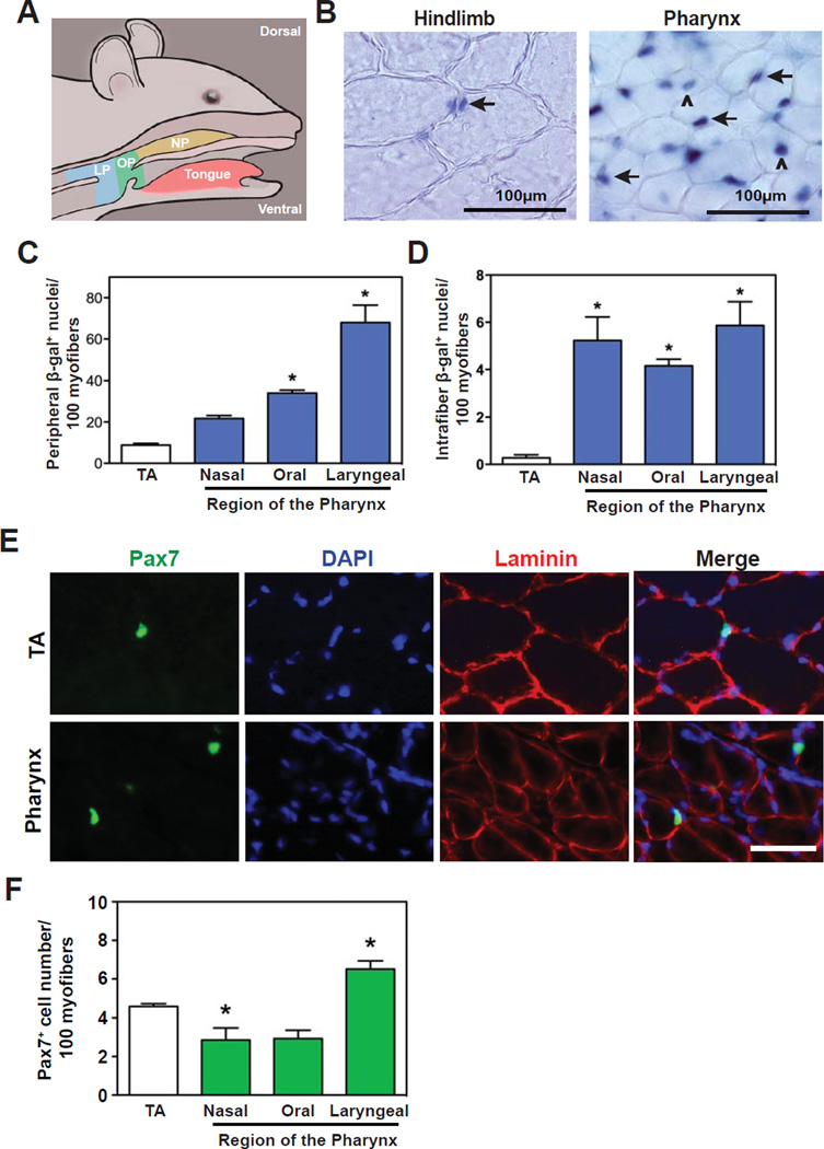
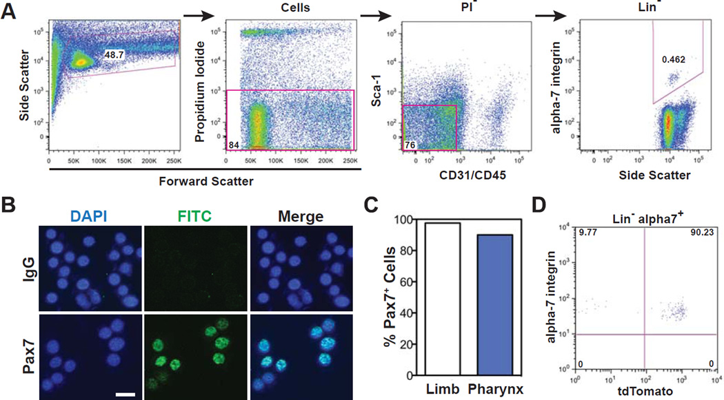
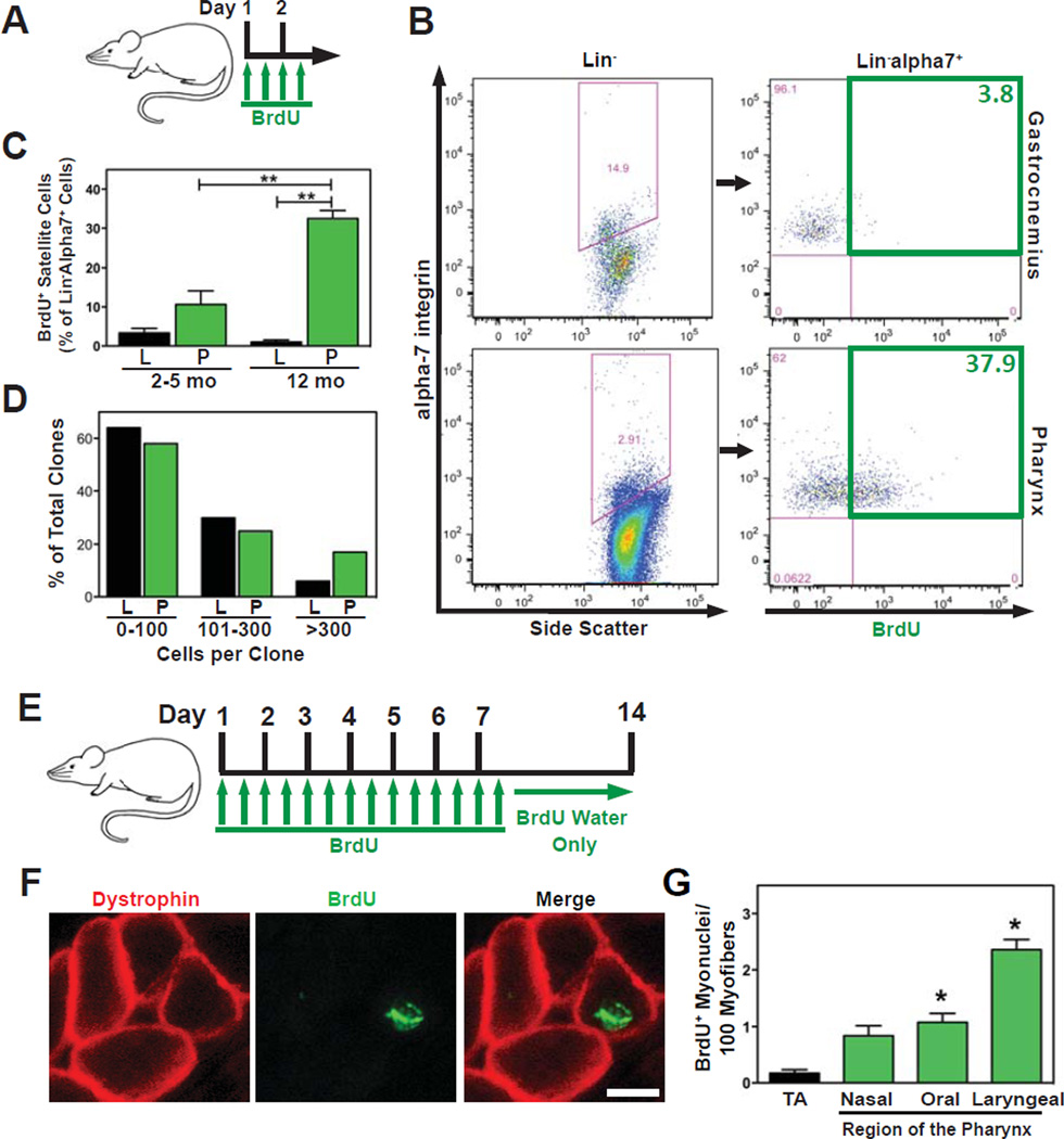
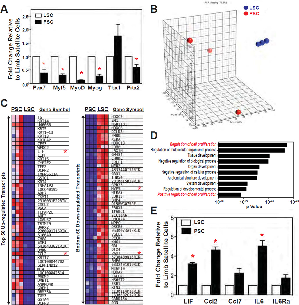
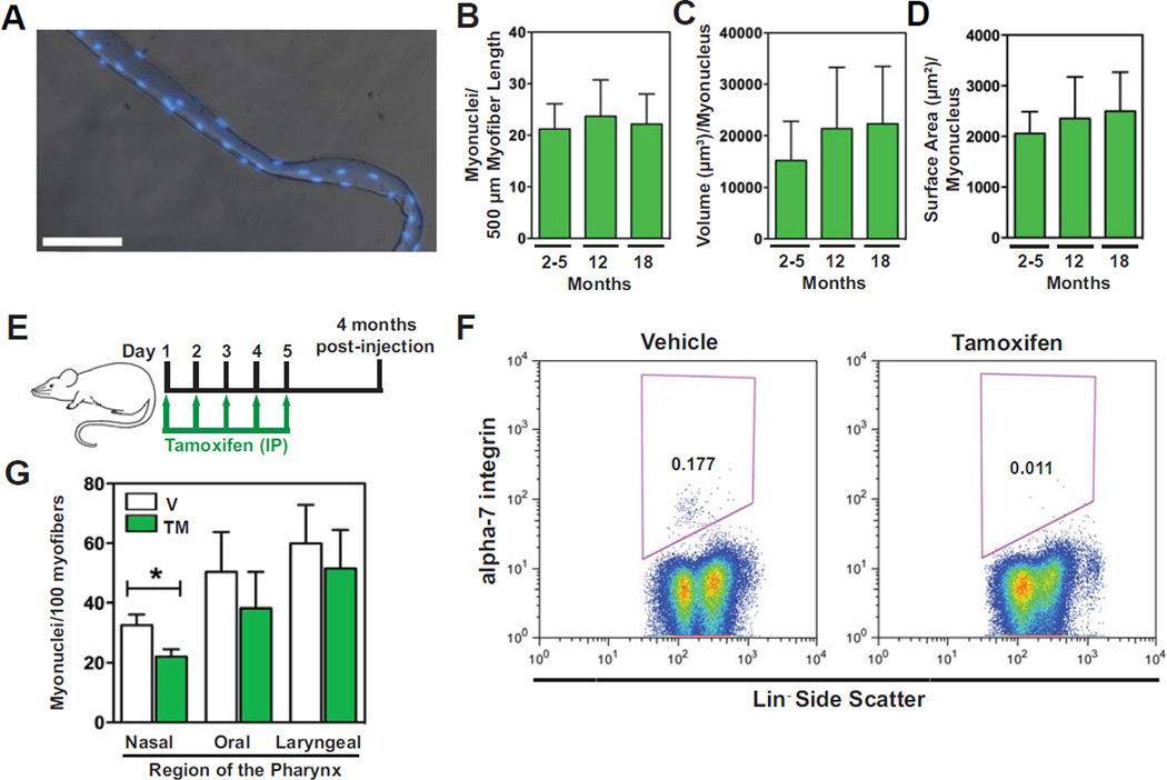
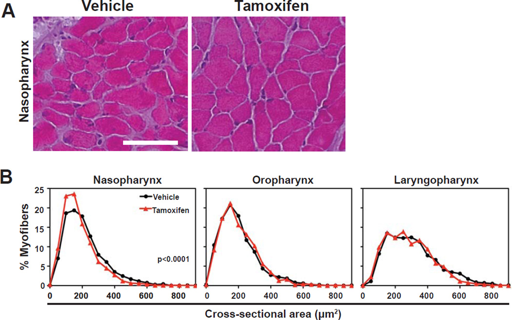
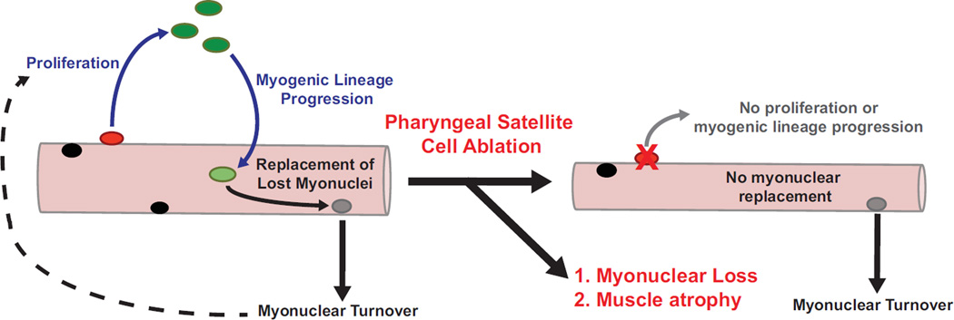
The pharyngeal muscles of the nasal, oral, and laryngeal pharynxes are required for swallowing. Pharyngeal muscles are preferentially affected in some muscular dystrophies yet spared in others. Muscle stem cells, called satellite cells, may be critical factors in the development of pharyngeal muscle disorders; however, very little is known about pharyngeal satellite cells (PSC) and their role in pharyngeal muscles. We show that PSC are distinct from the commonly studied hindlimb satellite cells both transcriptionally and biologically. Under basal conditions PSC proliferate, progress through myogenesis, and fuse with pharyngeal myofibers. Furthermore, PSC exhibit biologic differences dependent on anatomic location in the pharynx. Importantly, PSC are required to maintain myofiber size and myonuclear number in pharyngeal myofibers. Together, these results demonstrate that PSC are critical for pharyngeal muscle maintenance and suggest that satellite cell impairment could contribute to pharyngeal muscle pathology associated with various muscular dystrophies and aging.

Keywords: Muscle maintenance; Myofiber; Myonuclear turnover; Pharynx; Satellite cells.


Conflict of interest statement

Disclosure of Potential Conflicts of Interest

The authors report no potential conflicts of interest.

Figures

Figure 1
Figure 1. Pharyngeal muscles contain a larger number of activated satellite cells than hindlimb muscle
(A) Schematic of murine pharyngeal regions: NP = nasal pharynx; OP = oral pharynx; LP = laryngopharynx. (Modified from Randolph et al., 2014.) (B) Hindlimb (tibialis anterior) and pharyngeal muscles were collected at nine weeks of age from Myf5-nlacZ mice, sectioned, and incubated with X-gal to identify β-gal+ nuclei (blue). Representative muscle sections from hindlimb and oral pharynx (palatopharyngeus) are shown. Two types of β-gal+ nuclei were observed: peripherally located to myofibers (arrows) or centrally located within myofibers (Λ). (C) Each pharyngeal region contained increased numbers of peripherally located β-gal+ nuclei (satellite cells) versus hindlimb muscle. (D) Increased numbers of centrally localized β-gal+ myonuclei in myofibers were also observed in each pharyngeal region relative to hindlimb muscle. Data in C and D represent the mean ± SEM. *p < 0.05. n=4 mice. (E,F) Representative muscle sections from 8–9 week old C57BL/6 mice immunostained for Pax7 (green) and laminin (red). Pax7+ cells colocalizing with DAPI and laminin were identified (E) and quantified (F). Bar =100µm. Data represent the mean ± SEM. *p < 0.05. n=4 mice. TA= tibialis anterior muscle.
Figure 2
Figure 2. Identification of pharyngeal satellite cells using established cellular markers for hindlimb satellite cells
(A) Flow cytometry gating to identify and sort satellite cells isolated from pharyngeal muscle. Pharyngeal satellite cells were identified as α7-Integrin+ cells not derived from endothelial (CD31), hematopoietic (CD45), or fibro-adipogenic progenitor (Sca1) lineages (Lin α7-Integrin+). (B,C) Lin α7-Integrin+ cells were sorted and Pax7 expression visualized (B) and quantitated (C) in vitro using immunofluorescence. n= 509 pooled cells from 2 experiments of 10 mice each. (D) Approximately 90% of pharyngeal Lin α7-Integrin+ cells were tdTomato+ following tamoxifen-treatment of Pax7CreERTM/RosatdTomato heterozyotes. n=2 experiments of 2–3 mice pooled. PI = propidium iodide. Bar=20 µm.
Figure 3
Figure 3. Pharyngeal satellite cells proliferate and fuse with pharyngeal myofibers in the absence of induced injury
(A) Schematic of BrdU treatment protocol. (B) PI CD31 CD45 Sca1 α7-Integrin+ (Lin α7-Integrin+) satellite cells were identified using flow cytometry (left column) and analyzed to determine the percentage of proliferating satellite cells (BrdU+) in each tissue (right column). n=3 mice pooled. (C) Quantification of BrdU+ Lin α7-Integrin+ cells demonstrated a significantly larger proliferating population of satellite cells in pharyngeal versus hindlimb (gastrocnemius) muscles. Data represent the mean ± SEM. **p < 0.0001, n=3 experiments, 3–5 mice per experiment. (D) Lin α7-Integrin+ cells from either hindlimb (gastrocnemius/quadriceps) or pharyngeal muscles were sorted, plated at clonal densities and cultured for 8 days. Cultures were then fixed and stained with hematoxylin for quantification of cell number per clone. The number of large clones (>300 cells) was increased three-fold in pharyngeal versus hindlimb cultures. n=264 clones from 2 experiments using 10 mice each. (E) Schematic of BrdU treatment protocol. (F) Muscle sections were immunostained for dystrophin (red) and BrdU (green). BrdU+ nuclei contained within a dystrophin+ myofiber outline represent satellite cells that recently proliferated and fused into myofibers. Bar=50 µm. (G) Satellite cell fusion was quantified as the number of intrafiber BrdU+ nuclei per 100 myofibers. Satellite cell fusion occurred with higher frequency in pharyngeal muscles compared to hindlimb muscles. *p < 0.05, n=4 mice. L=hindlimb. P=pharynx. TA=tibialis anterior.
Figure 4
Figure 4. Comparative transcriptome analyses reveal pharyngeal and hindlimb satellite cells are distinct
Gene-expression analyses of FACS sorted pharyngeal and hindlimb satellite cells as determined by qRT-PCR (A,E) and microarray (B–D). (A) Selected regulatory transcripts involved in myogenesis were analyzed via qRT-PCR using RNA isolated from FACS sorted PSC and LSC. Data represent the mean fold-change of transcript steady-state levels ± SEM. n=3 experiments each containing 150,000–200,000 satellite cells pooled from 10–30 mice. *P<0.05. (B) Principal component analysis (PCA) of pharyngeal satellite cells (PSC, red dots) versus hindlimb (gastrocnemius/quadriceps) satellite cells (LSC, blue dots). PCA coordinates (PC1, 29.2%; PC2, 22.6%; and PC3, 18.4%) revealed a total data variation of 70.2%. n=3 experiments each containing 200,000 satellite cells pooled from 10–30 mice. (C) Heat maps comparing the levels of the top 50 transcripts either up- or down-regulated in PSC relative to LSC. Steady-state RNA levels are represented with a linear color scale ranging from dark red (enriched) to dark blue (depleted). Transcripts marked with red asterisks were validated by qRT-PCR. (D) Gene ontology (GO) process networks enriched in PSC generated with MetaCore Genego software. GO networks related to cell proliferation are highlighted in red. (E) qRT-PCR was used to validate microarray data of selected cytokine/chemokine transcripts that were enriched in PSC relative to LSC. Data represent the mean fold-change of transcript steady-state levels ± SEM. n=3 hindlimb and 4–5 pharyngeal experiments each containing 1500,000–200,000 satellite cells pooled from 10–30 mice. *P<0.05.
Figure 5
Figure 5. Myonuclear turnover occurs in pharyngeal muscle under basal conditions
(A) Merged DAPI and phase contrast images of a representative myofiber isolated from pharyngeal muscles. Bar=50µm. (B–D) Quantification of various pharyngeal myonuclear parameters indicated no change in myonuclear numbers with age. n=26–35 fibers per timepoint. (E) Schematic of satellite cell-specific ablation in Pax7CreERTM/RosaDTA-176 heterozygous mice. (F) Pharyngeal Lin α7-Integrin+ cells were ablated following tamoxifen-treatment of Pax7CreERTM/RosaDTA-176 (DTA/Pax7CreERTM) heterozygotes. Ablation efficiencies for pharyngeal Lin α7-Integrin+ cells ranged from 87–97%. n=8 experiments of 2–3 mice pooled. (G) Quantification of DAPI+ nuclei contained within dystrophin+ myofiber outlines revealed myonuclear loss within satellite cell-ablated muscles of the nasal pharynx. *p<0.05, n=3–4 mice per condition.
Figure 6
Figure 6. Pharyngeal satellite cells are required to prevent muscle atrophy in the nasal and laryngeal pharynxes
DTA/Pax7CreERTM mice received either vehicle or tamoxifen injections as described in Fig. 5E with pharyngeal muscles collected 4-months post-treatment. (A) Histologic sections of vehicle and tamoxifen treated mice. Bar=50µm. (B) Frequency distribution plots of myofiber cross-sectional areas from the naso-, oro-, and laryngopharyngeal regions are shown. Myofiber size of nasal pharyneal muscles decreased following satellite cell ablation. n=929–1505 myofibers, 3 mice per condition.
Figure 7
Figure 7. Model of basal pharyngeal satellite cell biology and maintenance of myofiber size
Pharyngeal satellite cells (red) proliferate, progress through myogenesis, and contribute myonuclei (black) to pharyngeal myofibers under basal conditions. The continual contribution of new myonuclei (light green) to pharyngeal myofibers counteracts the basal myonuclear loss (grey), preventing both loss of myonuclear numbers and myofiber size. Pharyngeal satellite cell impairment reduces myonuclear addition to pharyngeal myofibers resulting in both myonuclear loss and decreased myofiber size.

Similar articles

Cited by

References

    1. Hoffman EP, Brown RH, Jr, Kunkel LM. Dystrophin: the protein product of the Duchenne muscular dystrophy locus. Cell. 1987;51:919–928. - PubMed
    1. Monaco AP, Bertelson CJ, Liechti-Gallati S, et al. An explanation for the phenotypic differences between patients bearing partial deletions of the DMD locus. Genomics. 1988;2:90–95. - PubMed
    1. Nonaka I. Distal myopathies. Current opinion in neurology. 1999;12:493–499. - PubMed
    1. Bione S, Maestrini E, Rivella S, et al. Identification of a novel X-linked gene responsible for Emery-Dreifuss muscular dystrophy. Nat Genet. 1994;8:323–327. - PubMed
    1. Bonne G, Di Barletta MR, Varnous S, et al. Mutations in the gene encoding lamin A/C cause autosomal dominant Emery-Dreifuss muscular dystrophy. Nat Genet. 1999;21:285–288. - PubMed

Publication types