. 2015 Jul 16:10:146.
doi: 10.1186/s13014-015-0455-z.

Histone deacetylase inhibition sensitizes osteosarcoma to heavy ion radiotherapy

Affiliations

Histone deacetylase inhibition sensitizes osteosarcoma to heavy ion radiotherapy

Claudia Blattmann et al. Radiat Oncol. .

Abstract

Background: Minimal improvements in treatment or survival of patients with osteosarcoma have been achieved during the last three decades. Especially in the case of incomplete tumor resection, prognosis remains poor. Heavy ion radiotherapy (HIT) and modern anticancer drugs like histone deacetylase inhibitors (HDACi) have shown promising effects in osteosarcoma in vitro. In this study, we tested the effect of HIT and the combination of HIT and the HDACi suberoylanilide hydroxamic acid (SAHA) in a xenograft mouse model.

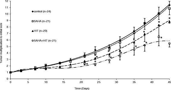
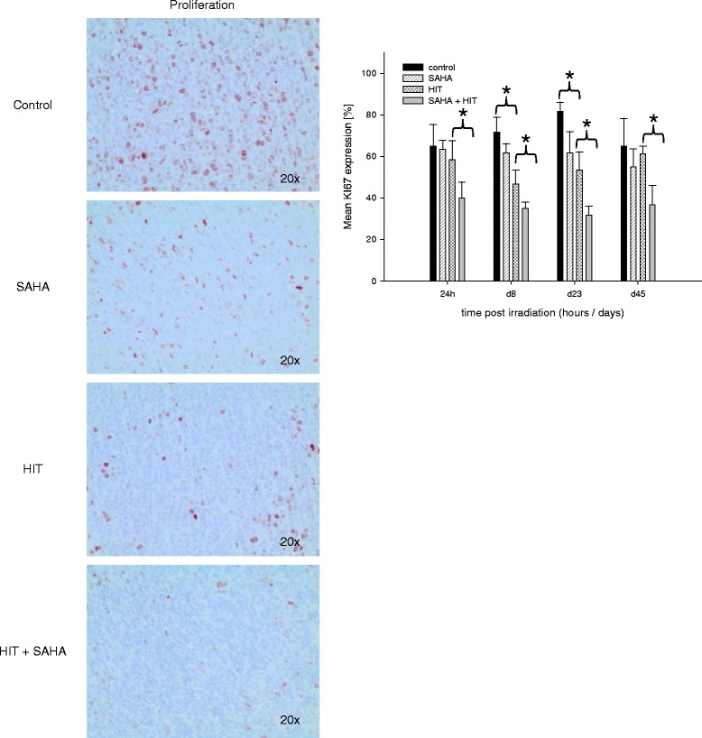
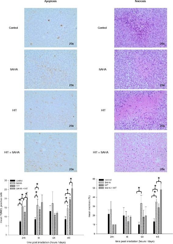
Methods: Osteosarcoma xenografts were established by subcutaneous injection of KHOS-24OS cells and treated with either vehicle (DMSO), SAHA, HIT or HIT and SAHA. Tumor growth was determined and tumor necrosis, proliferation rate, apoptotic rate as well as vessel density were evaluated.

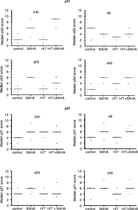
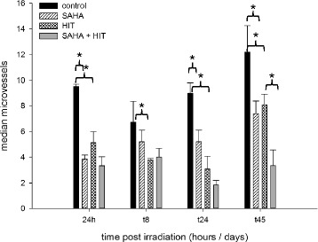
Results: Here, we show that the combination of HIT and SAHA induced a significant delay of tumor growth through increased rate of apoptosis, increased expression of p53 and p21(Waf1/Cip1), inhibition of proliferation and angiogenesis compared to tumors treated with HIT only.

Conclusion: HIT and in particular the combination of HIT and histone deacetylase inhibition is a promising treatment strategy in OS and may be tested in clinical trials.


Figures

Fig. 1
Fig. 1
HIT is superior to XRT in osteosarcoma xenografts. Tumor duplication after treatment with different doses of HIT and XRT was compared in to animal groups. Actual tumor growth delay was calculated with (T’x-Tx)/Tx as the time taken for the irradiated tumors (T’) and the control tumors (T) to x-fold multiply their volume (x)
Fig. 2
Fig. 2
The HIT dosage of 2.5 Gy leads to a tumor growth delay of 14 days. In a preliminary test, xenografts were irradiated with a single dose of 1, 2, 3, 5, 10 or 20 Gy. The dose of 2.5 Gy were chosen for our further experiments because significant but not to effective growth retardation within the first 14 days after treatment was seen leaving capacity for the anticipated additional effect of SAHA
Fig. 3
Fig. 3
The combination of HIT and SAHA results in a significant tumor growth delay compared to treatment with HIT or SAHA only. Osteosarcoma xenografts were treated with DMSO (controls), suberoylanilide hydroxamic acid (SAHA), irradiation (HIT) or SAHA plus HIT and tumor growth was determined until day 45 after HIT. Comparing HIT as mono-treatment to SAHA only, HIT seemed to be superior from day 10 on after treatment start reaching significance at day 23. The combination of HIT and SAHA yielded a significant (*) tumor growth retardation compared to SAHA only and HIT only starting day 20 and day 25 respectively
Fig. 4
Fig. 4
Combination of HIT and SAHA induces an increased local control. Tumor growth of osteosarcoma xenografts was determined after treatment with DMSO (controls), suberoylanilide hydroxamic acid (SAHA), irradiation (HIT) or SAHA plus HIT. Local control was defined as tumor growth > 1000 m3 and calculated according to the method of Kaplan and Meier. The combination of HIT and SAHA led to a significant local control compared to SAHA only and HIT only starting day
Fig. 5
Fig. 5
Treatment with HIT and SAHA results in a reduced proliferation in osteosarcoma xenografts. Histological analysis of proliferation in tumors treated with DMSO (control), suberoylanilide hydroxamic acid (SAHA), irradiation (HIT) or HIT and SAHA. The proliferation rate was significantly reduced in tumors after treatment with SAHA and HIT at all investigation time points. HIT only treatment led to a significantly lower proliferation rate 8 and 24 days after irradiation compared to the control group. SAHA only treatment had no significant effect on tumor proliferation
Fig. 6
Fig. 6
Impairment of apoptosis and necrosis after HIT and SAHA treatment. Apoptosis and necrosis in osteosarcoma xenografts were analyzed after treatment with DMSO (control), suberoylanilide hydroxamic acid (SAHA), irradiation (HIT) or HIT and SAHA. The combination treatment lead to a significant (*) induction of apoptosis one day, 8 and 45 days after HIT compared to HIT only treatment. Apoptosis was also increased after SAHA only and HIT only 24 h, 8 and 45 days after irradiation but not as much as treatment with HIT and SAHA. Combination of HIT and SAHA lead to a significant (*) induction of necrosis on day 45. SAHA only and HIT only treatment resulted in a significantly higher rate of necrosis from day 24 on but not at earlier time points compared to the control groups
Fig. 7
Fig. 7
Expression of p53 and p21WAF1/CIP1 is impaired in osteosarcoma xenografts after treatment with HIT and SAHA. Expression of p53 and p21WAF1/CIP1 was analyzed after treatment with DMSO (control), suberoylanilide hydroxamic acid (SAHA), irradiation (HIT) or HIT and SAHA. p53 expression was increased in all SAHA treated tumors compared to the vehicle treated control and only irradiated tumors on day 23 and 45. Increased p21Waf1/Cip1 expression was detected in tumors after SAHA only and SAHA and HIT treatment compared to the controls and the HIT treated tumors on day 8
Fig. 8
Fig. 8
Density of microvessels is significantly reduced in osteosarcoma after combination treatment. Xenografts were treated with DMSO (control), suberoylanilide hydroxamic acid (SAHA), irradiation (HIT) or HIT and SAHA. The density of microvessels was reduced in tumors of all treatment groups at all investigation time points. Tumors treated with HIT and SAHA showed a significant (*) lower vascularization compared to tumors treated with HIT from day 24 on and the lowest vascularization at all

References

    1. Schardt D, Elsässer T, Schulz-Ertner D. Heavy-ion tumor therapy: Physical and radio-biological benefits. Rev Mod Phys. 2010;82:383. doi: 10.1103/RevModPhys.82.383. - DOI
    1. Hundsdoerfer P, Albrecht M, Rühl U, Fengler R, Kulozik AE, Henze G. Long-term outcome after polychemotherapy and intensive local radiation therapy of high-grade osteosarcoma. Eur J Cancer. 2009;45(14):2447–51. doi: 10.1016/j.ejca.2009.06.006. - DOI - PubMed
    1. Blattmann C, Oertel S, Schulz-Ertner D, Rieken S, Haufe S, Ewerbeck V, et al. Non-randomized therapy trial to determine the safety and efficacy of heavy ion therapy in patients with non-resectable osteosarcomas. BMC Cancer. 2010;10:96. doi: 10.1186/1471-2407-10-96. - DOI - PMC - PubMed
    1. Sugahara S, Kamada T, Imai R, Tsuji H, Kameda N, Okada T, et al. Carbon ion radiotherapy for localized primary sarcoma of the extremities: results of a phase I/II trial. Working group for the bone and soft tissue sarcomas. Radiother Oncol. 2012;105(2):226–31. doi: 10.1016/j.radonc.2012.09.010. - DOI - PubMed
    1. Matsunobu A, Imai R, Kamada T, Imaizumi T, Tsuji H, Tsujii H, et al. Impact of carbon ion radiotherapy for unresectable osteosarcoma of the trunk. Working group for bone and soft tissue sarcomas. Cancer. 2012;118(18):4555–63. doi: 10.1002/cncr.27451. - DOI - PubMed

Publication types

MeSH terms